
Fig. 4
- ID
- ZDB-IMAGE-081001-2
- Genes
- Publication
- Majumdar et al., 2000 - Zebrafish no isthmus reveals a role for pax2.1 in tubule differentiation and patterning events in the pronephric primordia
- All Figures
- Figures for Majumdar et al., 2000
|
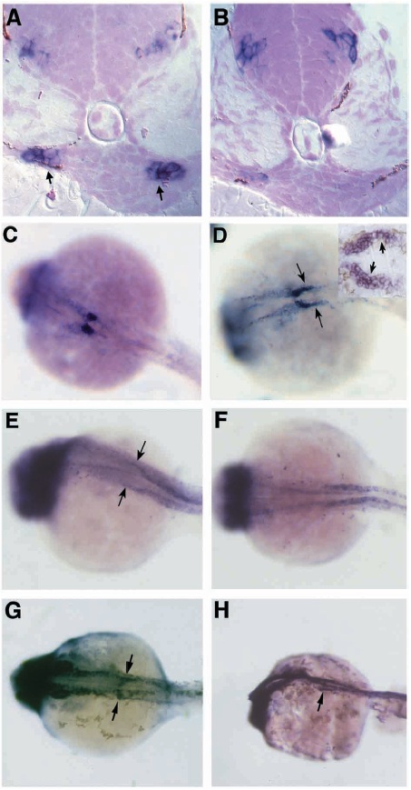
Fig. 4 Fig. 4. noi affects patterning of the nephric primordia. (A,C,E,G) Wildtype and (B,D,F,H) noitb21 embryos in situ hybridized with pax2.1 (A,B,G,H), wt1 (C,D) and a-subunit of Na+/K+ ATPase antisense probes (E,F) at 30 hpf (A,B,G,H) or 25 hpf (C-F). (A) Transverse histological sections (schematized by A in Fig. 3) of in situ hybridized 30 hpf embryos shows expression of pax2.1 in lateral, but not medial, cells of the pronephric primordia in wild type. (B) pax2.1 expression in nephric primordia is greatly reduced in noitb21 mutants, even though CNS expression still remains. (C) Whole-mount in situ hybridization with wt1 marks the nephric primordia in wild type. wt1 expression is caudally expanded in noitb21 (arrows in D). Sagittal section through a noitb21 pronephros stained with wt1 antisense probe (inset in D) reveals ectopic wt1 expression in lateral cells of the nephron primordia (arrows). Anterior duct expression of a-subunit of Na+/K+ ATPase (arrows in E) is lost in noitb21 embryos while more posterior duct expression remains (F). Whole-mount in situ hybridization with pax2.1 reveals tubule and duct expression in wild type (arrows in G). (H) Tubule expression is gone, but anterior duct pax2.1 expression remains in noitb21.