
Fig. 2
- ID
- ZDB-IMAGE-080923-2
- Genes
- Publication
- Tallafuss et al., 2008 - The Met receptor tyrosine kinase prevents zebrafish primary motoneurons from expressing an incorrect neurotransmitter
- All Figures
- Figures for Tallafuss et al., 2008
|
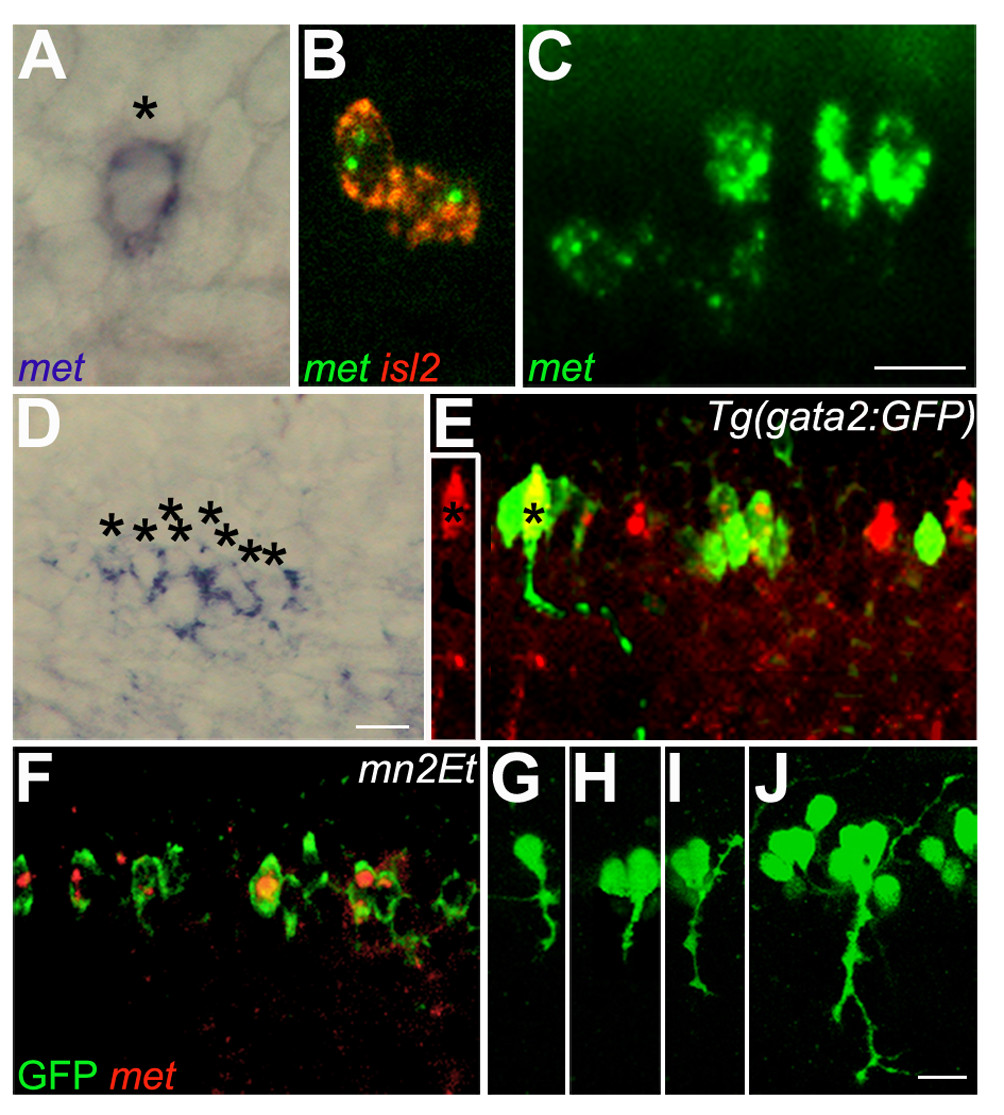
Fig. 2 Zebrafish met is expressed in developing spinal motoneurons. All photographs are dorsal to the top and anterior to the left; this is also the case for subsequent figures except where noted. (a) A 22 hpf embryo showing met RNA expression in one cell of a spinal hemisegment (asterisk). (b) A 22 hpf embryo showing co-expression of met (green) and islet2 (red) in CaP and VaP. (c) A 26 hpf embryo showing met expression in all four PMNs in this spinal hemisegment. (d) A 48 hpf embryo showing met expression in eight cells (asterisks) in this hemisegment. (e) A 48 hpf gata2:GFP transgenic embryo showing GFP expression in ventrally projecting SMNs (green) and met expression (red) in a subset of these cells. The axon of the SMN labeled with an asterisk is shown as it projects out of the spinal cord toward its ventral muscle target. The inset to the left shows the same SMN, also marked with an asterisk, in only the red channel, clearly revealing that the SMN expresses met RNA. (f) A 48 hpf mn2Et transgenic embryo showing GFP expression (green) in PMNs and SMNs; met (red) is expressed in a subset of these cells. (g-j) mn2Et transgenic embryos at 24 hpf showing GFP expression in motoneurons: posterior segment showing GFP expression in CaP (g); posterior segment showing GFP expression in CaP and VaP (h); a more anterior segment showing GFP expression in CaP and MiP (i); an even more anterior segment showing GFP expression in CaP, VaP, MiP, RoP and several SMNs (j). Note that even as late as 5 days post-fertilization, in mn2Et embryos GFP-positive cells in the spinal cord all appear to have peripheral axons and no interneuron-like cells express GFP, suggesting that in the mn2Et line GFP is expressed exclusively in motoneurons. Scale bars, 10 μm.