
Fig. 7
- ID
- ZDB-IMAGE-080917-1465
- Genes
- Antibodies
- Publication
- Piotrowski et al., 2000 - The endoderm plays an important role in patterning the segmented pharyngeal region in zebrafish (Danio rerio)
- All Figures
- Figures for Piotrowski et al., 2000
|
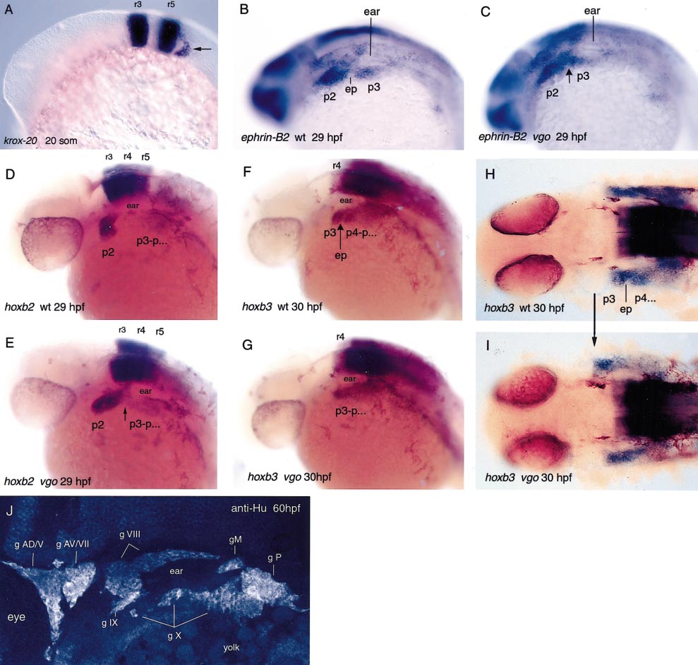
Fig. 7 In situ hybridization with krox-20 (A), ephrin-B2 (B, C), hoxb2 (D, E), and hoxb3 (F–I), which are segmentally expressed in the hindbrain. In 20-somite-old vgo mutant embryos krox-20 is normally expressed in rhombomeres 3 and 5 (A), as well as in the neural crest cells that migrate from rhombomere 5 into pharyngeal arches 3–7 posterior to the ear (arrow). These cells contribute to the third neural crest stream. At this stage the posterior endodermal pouches have not formed yet and therefore the pharyngeal arches 3–7 cannot be distinguished. The expression patterns of hoxb2 in r3, r4, and r5 in 29-hpf wild-type (D) and vgo embryos (E) are indistinguishable from each other, indicating that hindbrain segmentation is not affected. However, in vgo (E) the neural crest cells that have migrated from r4 and r7 into the second (p2) and more posterior arches spread anteriorly along the A-P axis. In the pharyngeal arch region, r7-derived neural crest cells contact r4-derived neural crest cells (arrow), indicating that the arches are not separated from each other. hoxb3 is expressed in the third neural crest stream that migrates into p3 and the more posterior arches (F, H). No endodermal pouches form within the hoxb3 expression domain in vgo (F, G, arrow; ep, endodermal pouch). The expression is anteriorly expanded in vgo and is adjacent to the posterior expression boundary of hoxb2 in p2 (H, I, arrow). The sharp anterior expression boundary of hoxb3 suggests that neural crest cells from p2 and p3 do not intermingle (I). The sensory cranial ganglia develop normally in 60-hpf vgo as visualized with an antibody against Hu proteins that are expressed in neurofilaments (J). Sensory cranial ganglia possess a neural crest contribution, suggesting that in vgo the neural crest cells are correctly specified and migrate to their proper targets. The nomenclature of Raible and Kruse (2000) and Piotrowski and Northcutt (1996) was adopted. Abbreviations: g AD, anterodorsal lateral line ganglion; g AV, anteroventral lateral line ganglion; g M, middle lateral line ganglion; g P, posterior lateral line ganglion; g V, trigeminal ganglion; g VII, facial sensory ganglion; g VIII, octaval ganglion; g X, vagus ganglion.
Reprinted from Developmental Biology, 225(2), Piotrowski, T. and Nüsslein-Volhard, C., The endoderm plays an important role in patterning the segmented pharyngeal region in zebrafish (Danio rerio), 339-356, Copyright (2000) with permission from Elsevier. Full text @ Dev. Biol.