
Fig. 3
- ID
- ZDB-IMAGE-080908-13
- Genes
- Publication
- Fish et al., 2008 - miR-126 regulates angiogenic signaling and vascular integrity
- All Figures
- Figures for Fish et al., 2008
|
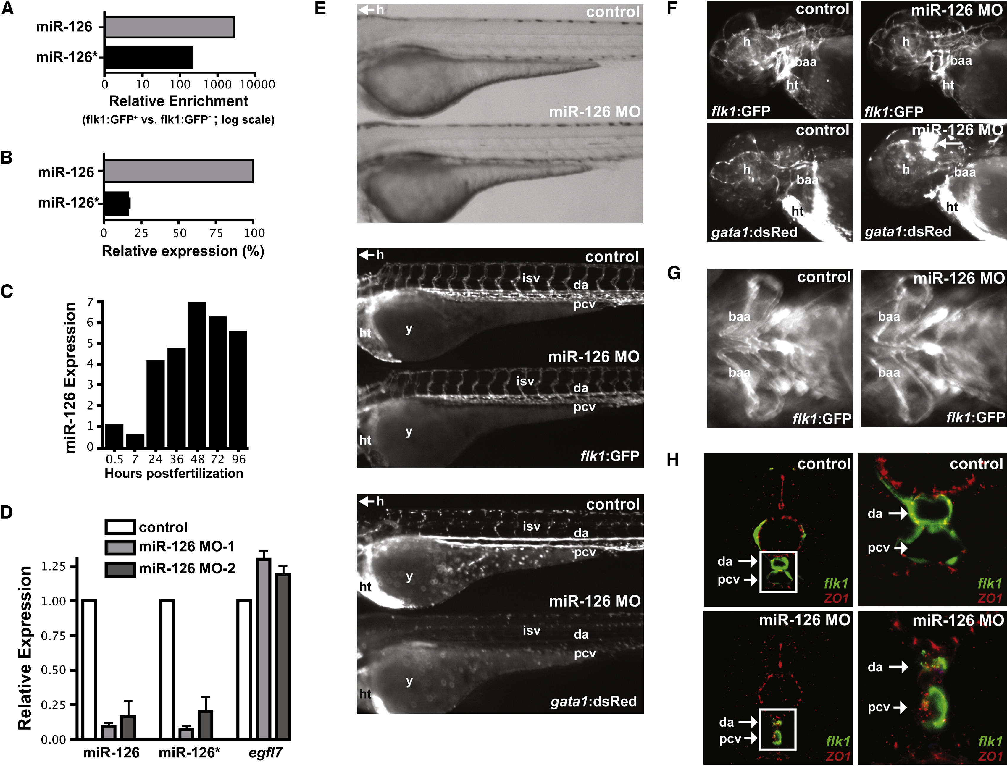
Fig. 3 miR-126 Regulates Vascular Integrity and Lumen Maintenance In Vivo
(A) miR-126 and miR-126* enrichment (qRT-PCR) in GFP+ endothelial cells from 72 hpf Tg(flk1:GFP)s843 zebrafish compared to GFP- cells.
(B) Relative levels of miR-126 and miR-126* in 72 hpf zebrafish embryos.
(C) miR-126 expression monitored by qRT-PCR during zebrafish development.
(D) Levels of miR-126/126* or egfl7 (across intron containing miR-126) quantified by qRT-PCR in 72 hpf zebrafish injected with miR-126 MOs relative to control.
(E) Lateral views of control and miR-126 MO-injected Tg(flk1:GFP)s843; Tg(gata1:dsRed)sd2 zebrafish (72 hpf). Brightfield microscopy (top) revealed no major changes in gross morphology, while flk1:GFP showed normal blood vessel patterning (middle). Presence of blood cells (gata1:dsRed) in the intersomitic vessels (isv), dorsal aorta (da) and primary cardinal vein (pcv) was greatly reduced in morphants (bottom). y, yolksac; h, head; ht, heart.
(F) miR-126 morphants (48 hpf) had normal vessel patterning (flk1:GFP), but developed cranial hemorrhages (gata1:dsRed; arrow) in the head. baa, branchial arch arteries.
(G) Ventral view of baa suggested smaller luminal size in miR-126 morphants.
(H) Transverse section of control or miR-126 MO-treated zebrafish revealed that the da and pcv of morphants had a smaller lumen than controls; higher magnification (right panels) of boxed area shows collapsed da and small pcv in morphants. flk1:GFP, green; ZO-1, an epithelial marker, red.
Reprinted from Developmental Cell, 15(2), Fish, J.E., Santoro, M.M., Morton, S.U., Yu, S., Yeh, R.F., Wythe, J.D., Ivey, K.N., Bruneau, B.G., Stainier, D.Y., and Srivastava, D., miR-126 regulates angiogenic signaling and vascular integrity, 272-284, Copyright (2008) with permission from Elsevier. Full text @ Dev. Cell