
IMAGE
Fig. S3
- ID
- ZDB-IMAGE-080828-49
- Genes
- Publication
- Lokmane et al., 2008 - Crucial role of vHNF1 in vertebrate hepatic specification
- All Figures
- Figures for Lokmane et al., 2008
Image
|
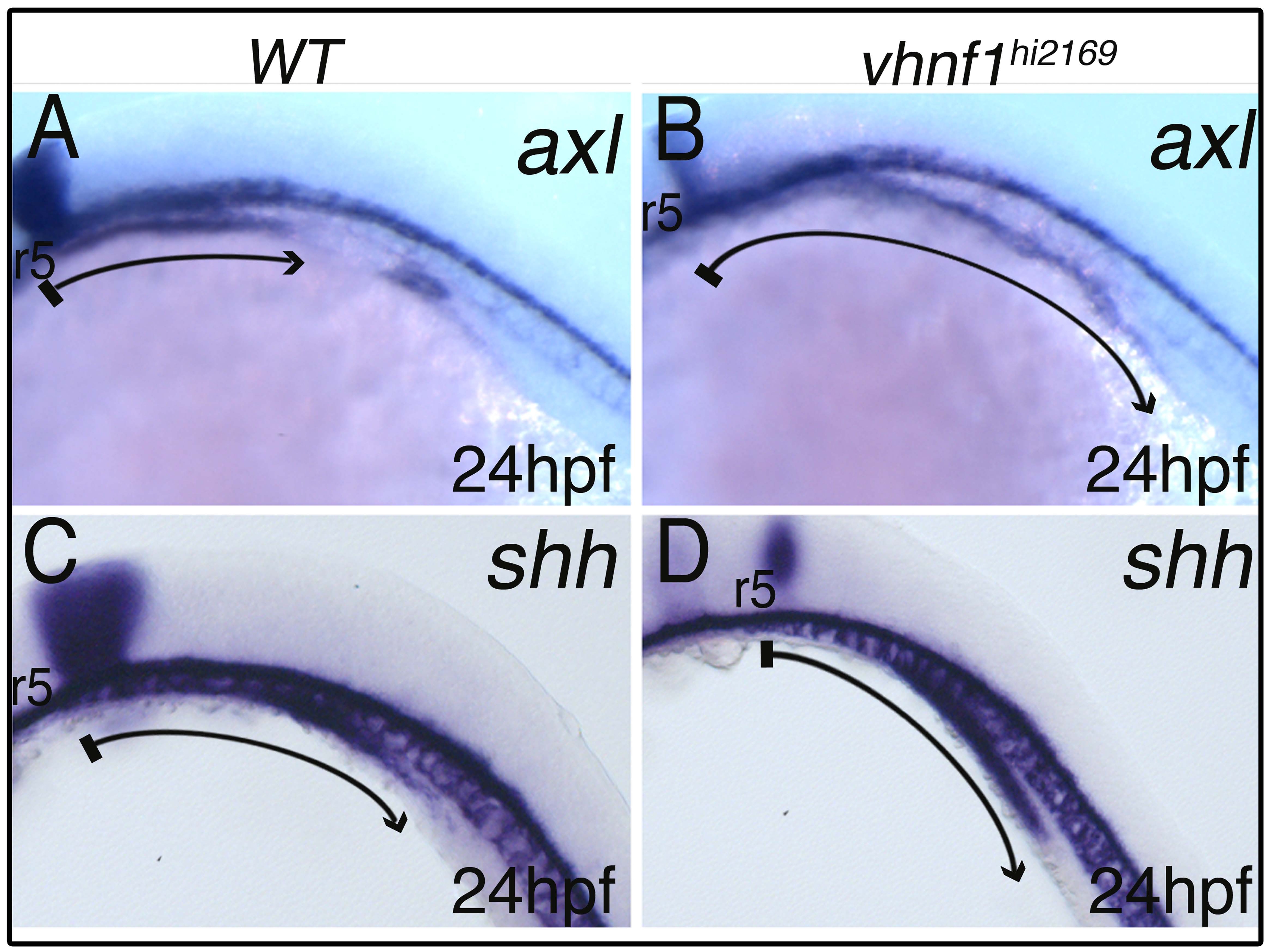
Figure Caption
Fig. S3 Defective gut regionalisation in the vhnf1hi2169 mutant. Whole-mount in situ hybridization in lateral view at 24 hpf. Anterior to the left. krox20 (egr2b) or frb35 (egr2a) staining was used to identify rhombomere 5 (r5), which is strongly reduced in vhnf1hi2169 homozygous embryos. axial (foxa2) and shh are caudally expanded in the mutant gut (B,D) as compared with the wild type (A,C).
Figure Data
Acknowledgments
This image is the copyrighted work of the attributed author or publisher, and
ZFIN has permission only to display this image to its users.
Additional permissions should be obtained from the applicable author or publisher of the image.
Full text @ Development