
Fig. 3
- ID
- ZDB-IMAGE-080723-2
- Genes
- Publication
- Boisset et al., 2008 - Characterization of pip5k3 fleck corneal dystrophy-linked gene in zebrafish
- All Figures
- Figures for Boisset et al., 2008
|
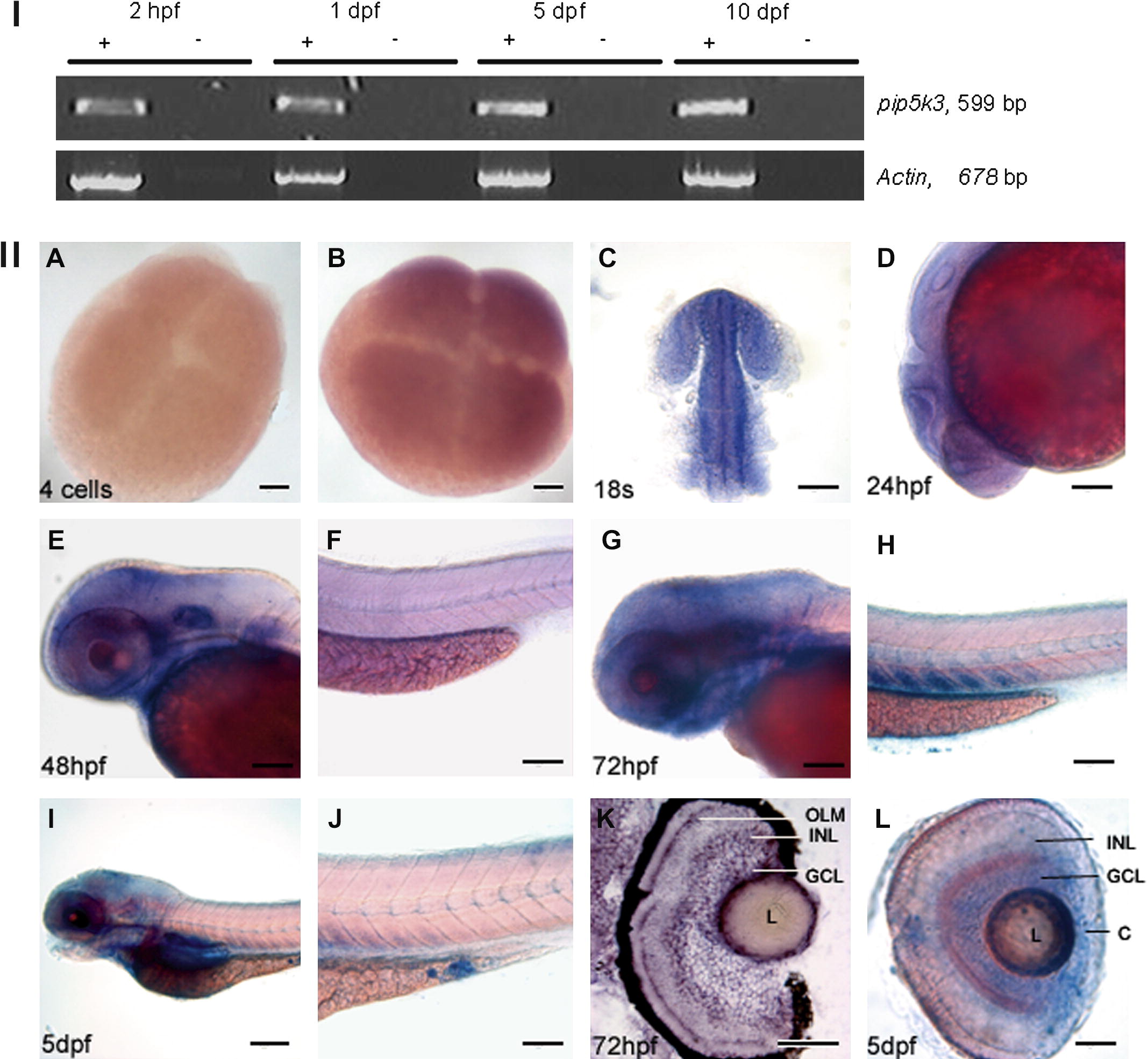
Fig. 3 Embryonic expression of zebrafish pip5k3. (I) RT-PCR analysis of embryonic pip5k3 during development. Pip5k3 transcripts were detected at 2 hpf, 1 dpf, 5 dpf and 10 dpf. Absence of DNA contamination was controlled by a no reverse transcriptase experiment. The amplified fragments lengths are written on the right. (II) Whole mount in situ hybridization. (A) Control experiments using sense probe. (B, C, dorsal view and D, lateral view) pip5k3 was uniformly expressed at 4-cell stage, 18 somites and 24 hpf. (E, G and I) Expression of pip5k3 in the head region at 48 hpf, 72 hpf and 5 dpf. Note that the staining in the gut region was not specific compared to the sense probe (I and data not shown). (F, H and J) View of the tail of 48 hpf, 72 hpf and 5 dpf embryos. No staining was detected at 48 hpf in contrast to 72 hpf and 5 dpf where a partial expression of pip5k3 was found in somites. Note that the blue spot localized to the proctodeum at 5 dpf was an artefact (J). (K) In situ on 12 μm cryosectionned eye of 3 dpf-old larva. Staining was mainly observed in OLM, INL, GCL and L. (L) Eye of 5 dpf-old larva showing expression of pip5k3 in INL, GCL, L and C. OLM, outer limiting membrane; INL, inner nuclear layer; GCL, ganglion cell layer; L, lens; C, cornea. Scale bars: (A and B) 20 μm; (C–H and J) 100 μm; (I) 200 μm; (K and L) 50 μm.
Reprinted from Gene expression patterns : GEP, 8(6), Boisset, G., Polok, B.K., and Schorderet, D.F., Characterization of pip5k3 fleck corneal dystrophy-linked gene in zebrafish, 404-410, Copyright (2008) with permission from Elsevier. Full text @ Gene Expr. Patterns