
Fig. 2
- ID
- ZDB-IMAGE-080723-1
- Genes
- Publication
- Boisset et al., 2008 - Characterization of pip5k3 fleck corneal dystrophy-linked gene in zebrafish
- All Figures
- Figures for Boisset et al., 2008
|
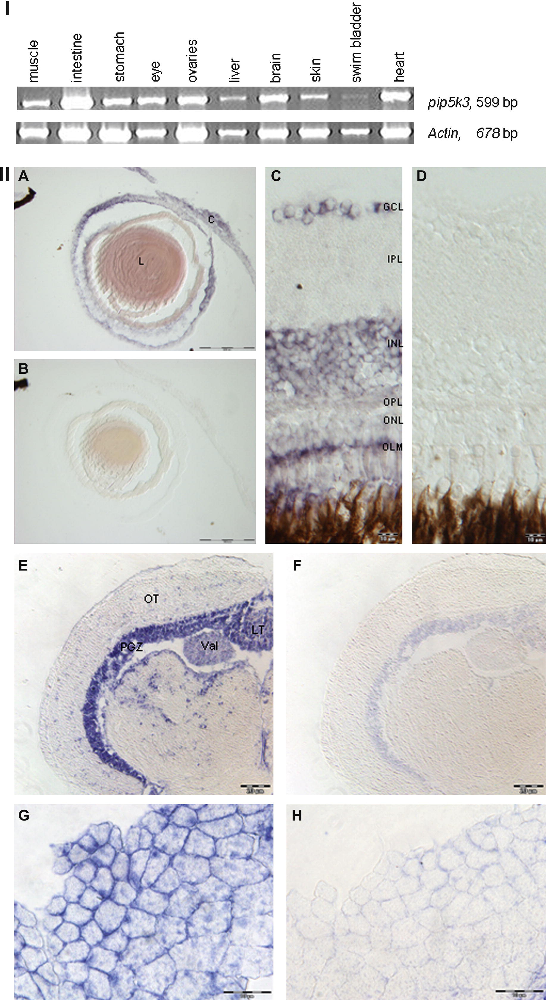
Fig. 2 Expression profile of adult zebrafish pip5k3. (I) RT-PCR analysis of pip5k3 expression in different tissues of zebrafish. Pip5k3 was expressed in all tested tissues, but at low level in swim bladder. The house-keeping gene actin was used as control. The size of the amplified fragments is indicated on the right. (II, A–H) In situ hybridization using RNA probe specific for zebrafish pip5k3 on adult eye, brain and muscle cryosections. (A) Pip5k3 was expressed in the cornea and lens. (C) In the retina, pip5k3 staining was seen in GCL, INL and OLM. (E) In the brain, pip5k3 staining was mainly observed in PGZ, Val and TL. (G) Pip5k3 staining was perinuclear and located to the membranes of the cells in the muscle. (B, D, F and H) Control staining with sense probe. C, cornea; L, lens; GCL, ganglion cell layer; INL, inner nuclear layer; IPL, inner plexiform layer; LT, longitudinal torus; OLM, outer limiting membrane; ONL, outer nuclear layer; OPL, outer plexiform layer; OT, optic tectum; PGZ, periventricular gray zone of the optic tectum; Val, lateral division of valvula cerebelli. Scale bars: (A and B) 100 μm; (C and D and G and H) 10 μm; (E and F) 20 μm.
Reprinted from Gene expression patterns : GEP, 8(6), Boisset, G., Polok, B.K., and Schorderet, D.F., Characterization of pip5k3 fleck corneal dystrophy-linked gene in zebrafish, 404-410, Copyright (2008) with permission from Elsevier. Full text @ Gene Expr. Patterns