
Fig. 8
|
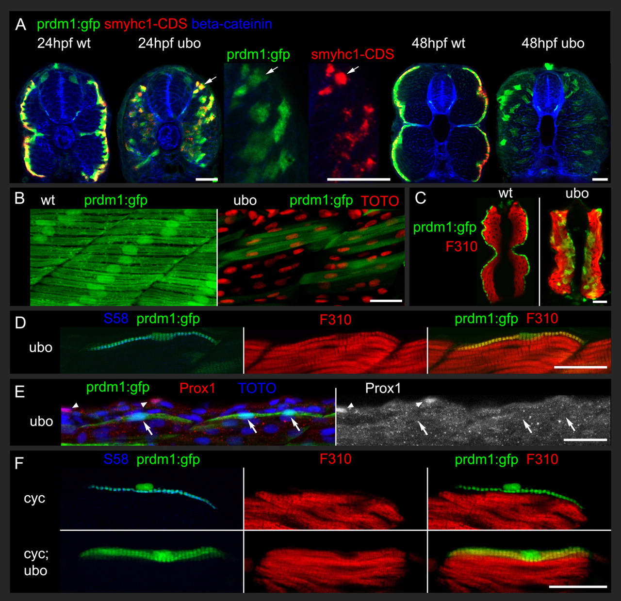
Fig. 8 The prdm1:gfp transgenic reporter provides a lineage tracer to follow the fate of prdm1-dependent muscle precursors in ubo (prdm1) mutants. (A) Transverse sections show colocalisation prdm1:gfp with smyhc1-CDS in situ hybridisation in ubo (prdm1)tp39 mutants and wild-type siblings at 24 hpf. At 48 hpf, the prdm1:gfp marked muscle fibres are still present in ubo (prdm1)tp39 mutants, although Smyhc expression is reduced. Cell boundaries are marked with anti-β-catenin 15B8. (B) Lateral views showing that muscle fibres with prdm1:gfp form a superficial layer of horizontal, mononucleate fibres in 48 hpf wild-type embryos but are among the fast fibres in ubo (prdm1)tp39 mutants and like fast fibres are frequently multinucleate (TOTO stain) and orientated diagonally. (C) Transverse sections at 48 hpf show colocalisation of F310 fast muscle antigen with prdm1:gfp in ubo (prdm1)tp39 mutants but not in wild-type siblings. (D) In posterior trunk at 48 hpf, in ubo (prdm1)tp39 mutants, the dorsal fibres with persistent slow MyHC S58 antigen are marked with prdm1:gfp and fast muscle F310 antigen. (E) In posterior trunk at 36 hpf, in ubo (prdm1)tp39 mutants, even the most dorsally positioned fibres marked with prdm1:gfp lack Prox1 (arrows). Note the intense Prox1 staining in isolated cells external to the myotome (arrowheads). (F) In posterior trunk of cyclopamine-treated embryos, prdm1:gfp marked secondary fibres colocalise with slow MyHC S58 antigen but not fast muscle F310 antigen in genotyped wild-type siblings and vice versa in genotyped ubo (prdm1)tp39 mutants. Scale bars: 25 μm.