
|
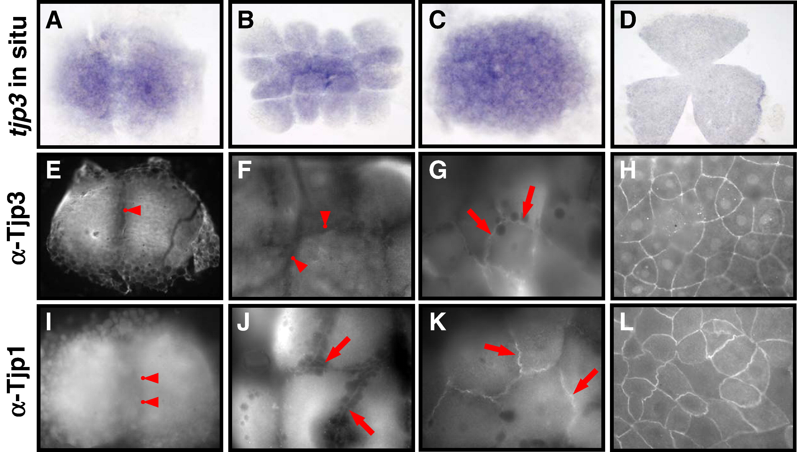
Fig. 1 Tight junction biogenesis in the early zebrafish embryo. Tjp3/zo-3 whole mount in situ hybridization (A–D) and Tjp3/Zo-3 (E–H) and Tjp1/Zo-1 whole-mount immunofluorescence (I–L) of 2-cell stage/0.75 h (A, E, I); 8–16-cell stage/1.25 hpf (B, F, J); 64-cell stage/2 hpf (C, G, K); shield stage/6 hpf (D, H, L). Tjp3/zo-3 is a maternally supplied mRNA with in situ hybridization signals from the 2-cell stage (A–C). Expression is uniform up to 60% epiboly (D). Tjp1/Zo-1 and Tjp3/Zo-3 are already present in the 2-cell stage embryo, where they are localized diffusely in the cytoplasm but not at cell–cell contact sites (E, anti-Tjp3/Zo-3; I, anti-Tjp1, arrowheads). Tjp3/Zo-3 remains absent from cell–cell contact sites at the 8-cell stage (F, arrowheads) but Tjp1/Zo-1 already accumulates at cell–cell junctions (J, arrows). Tjp3/Zo-3 gets localized to cell–cell contacts only at the 64-cell stage (G, arrows), by a time when Tjp1/Zo-1 is strongly present in the junctional complex (K, arrow). By 6&nbs accumulates at cell–cell junctions (J, arrows). Tjp3/Zo-3 gets localized to cell–cell contacts only at the 64-cell stage (G, arrows), by a time when Tjp1/Zo-1 is strongly present in the junctional complex (K, arrow). By 6 hpf the enveloping cell layer contains Tjp3/Zo-3 and Tjp1/Zo-1 at all cell–cell junctions (G, H).
Reprinted from Developmental Biology, 316(1), Kiener, T.K., Selptsova-Friedrich, I., and Hunziker, W., Tjp3/zo-3 is critical for epidermal barrier function in zebrafish embryos, 36-49, Copyright (2008) with permission from Elsevier. Full text @ Dev. Biol.