
|
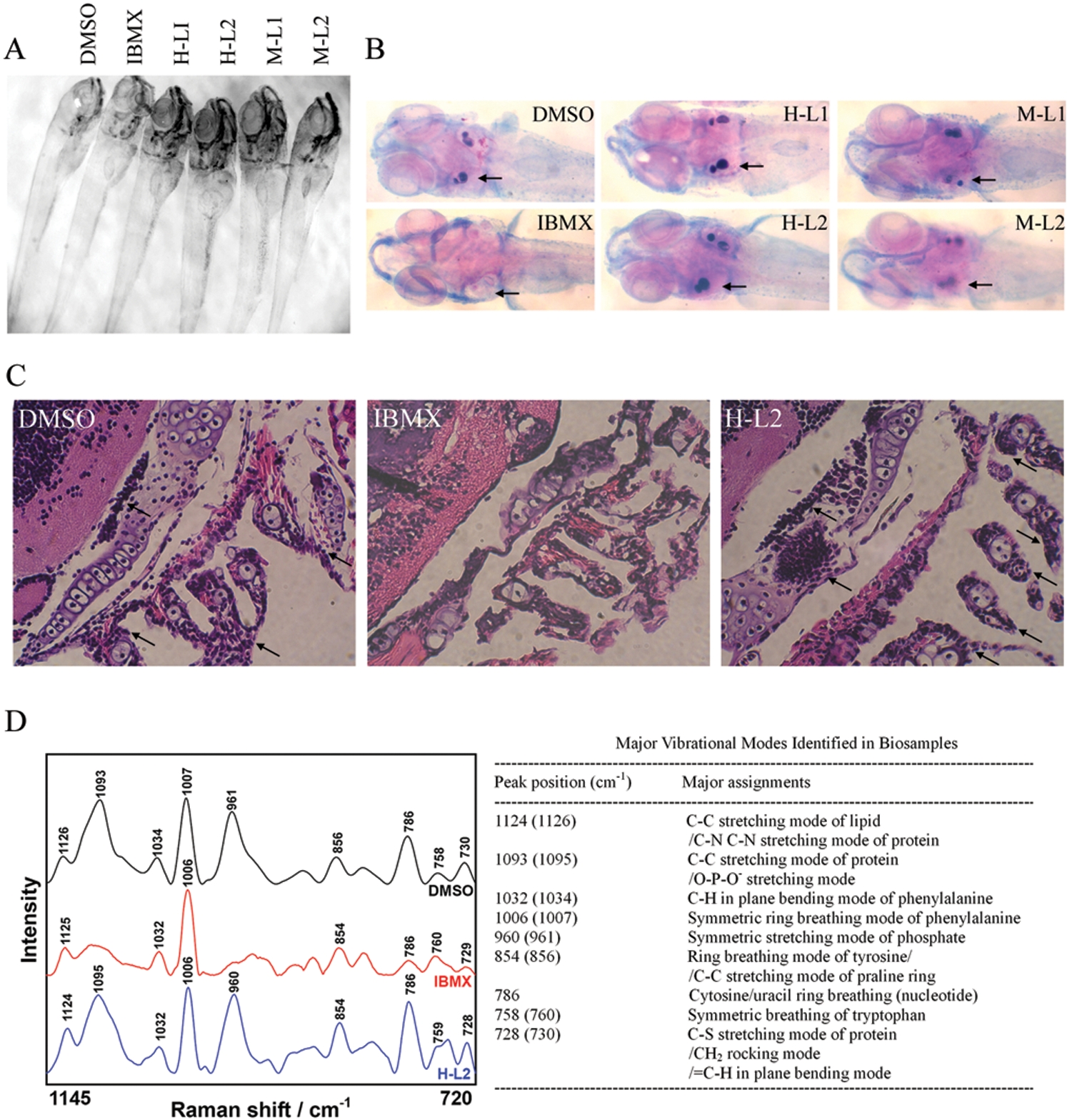
Fig. 5 Leptin abrogates IBMX-induced lack of skeletal ossification in developing zebrafish. Embryos were treated in vehicle DMSO, IBMX (0.045 mM), and IBMX with human [H] or mouse [M] leptin (L1: 0.6 μg/mL, L2: 1.5 μg/mL) at 2 dpf. (A) Morphology of embryos at 8 dpf. (B) Alizarin Red S and alcian blue staining of whole zebrafish reveals normal skeletal ossification in DMSO-treated embryos at otolith (arrow), while absent skeletal ossification is evident in IBMX-treated embryos. The addition of human and mouse leptin both in low and high concentrations abrogates IBMX-induced absence of skeletal ossification. (C) Hematoxylin-and-eosin staining of zebrafish sections reveals impaired bone development in IBMX-treated embryos. Arrows indicate bone development with cell aggregation at otolith and pharyngeal arches. (D) Raman microspectroscopic analysis of bone development at otolith in zebrafish.