
IMAGE
Fig. 4
- ID
- ZDB-IMAGE-080114-21
- Genes
- Publication
- Schneider et al., 2008 - Calcium fluxes in dorsal forerunner cells antagonize {beta}-catenin and alter left-right patterning
- All Figures
- Figures for Schneider et al., 2008
Image
|
Figure Caption
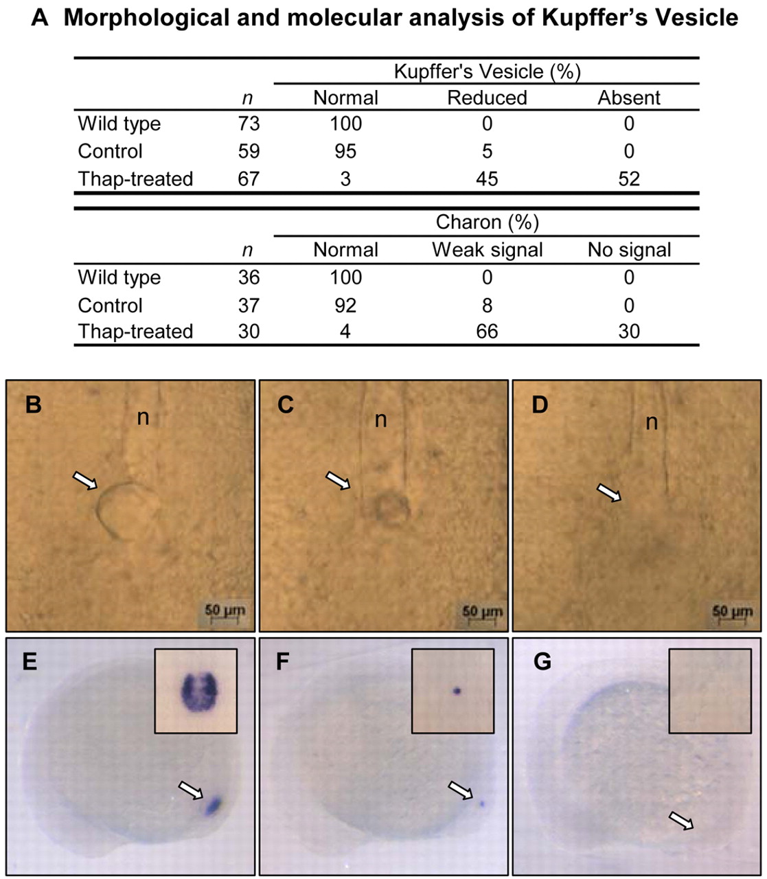
Fig. 4 DFC regional Ca2+ is required for KV morphogenesis. (A) Summary of KV defects. (B-D) Bright field image of wt embryo with normal KV (B) and thapsigargin-treated with reduced (C) and absent (D) KV: dorsal view, 10 somites. Arrows indicate vesicle location; n, notochord. (E-G) charon expression (arrows) around KV in wt (E) and thapsigargin-treated embryos with reduced (F) and absent (G) signal: lateral view, 10 somites. Insets show dorsal view of charon expression. Thap, thapsigargin. Scale bars: 50 μm.
Figure Data
Acknowledgments
This image is the copyrighted work of the attributed author or publisher, and
ZFIN has permission only to display this image to its users.
Additional permissions should be obtained from the applicable author or publisher of the image.
Full text @ Development