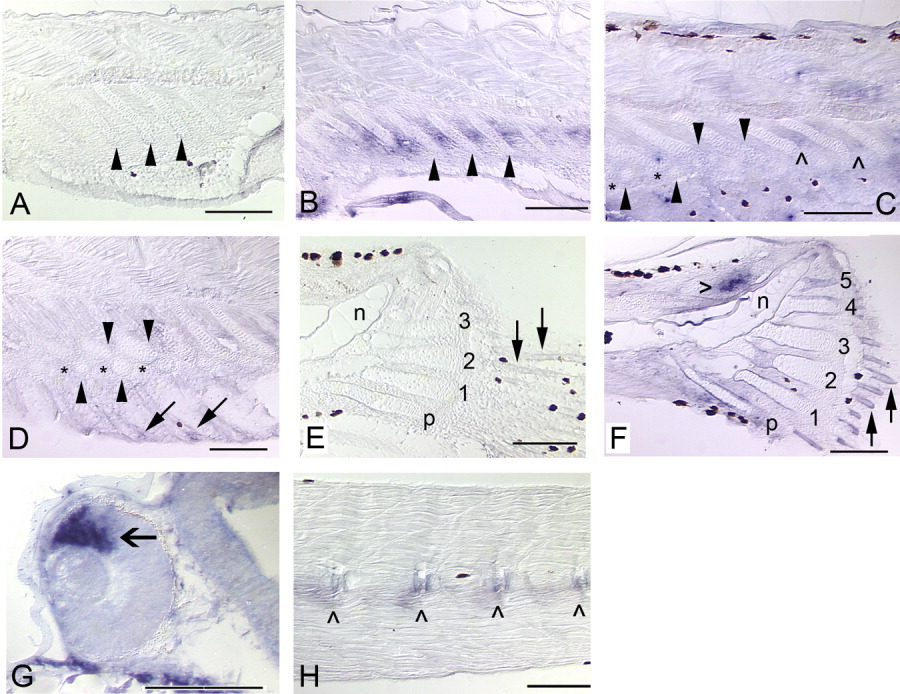
Fig. 5 Expression of bmp4 in the median fins and axial skeleton of zebrafish. Solid arrows mark fin rays, arrowheads mark radials, and hypurals are numbered. Black patches are melanophores. Anterior is to the left and dorsal to the top. A: In the same individual as H, bmp4 expression is not detected in the anal fin (5.4 mm). B: At a slightly later stage, bmp4 expression is observed in the interradial mesenchyme (5.7 mm). C: Expression of bmp4 is not detected in the ZS (*) between the proximal (top row of arrowheads) and distal (bottom row of arrowheads) radials, although it is maintained in the interradial mesenchyme (∧) between more posteriorly located, less-developed radials (6.0 mm). D: When radial segmentation is complete, expression of bmp4 is detected only in the fin rays (arrows; 6.6 mm). E: In the same individual as H, bmp4 expression is not detected in the caudal fin (5.4 mm). F: At a slightly later stage, expression is observed in the fin rays and ossifying perichondrium surrounding the hypurals, and very strongly in mesenchyme (hollow arrowhead) dorsal to the notochord (n) of the caudal fin (5.7 mm). G: An open arrow marks expression in the dorsal retina (Chin et al.,[1997]), which served as a positive control (26 hpf). H: Expression of bmp4 in the developing axial skeleton (centra, ∧) is not as robust as the dorsal retina positive control, but is clearly detectable (5.4 mm). Scale bars = 0.1 mm.
Image
Figure Caption
Figure Data
Acknowledgments
This image is the copyrighted work of the attributed author or publisher, and
ZFIN has permission only to display this image to its users.
Additional permissions should be obtained from the applicable author or publisher of the image.
Full text @ Dev. Dyn.

