
Image
|
Figure Caption
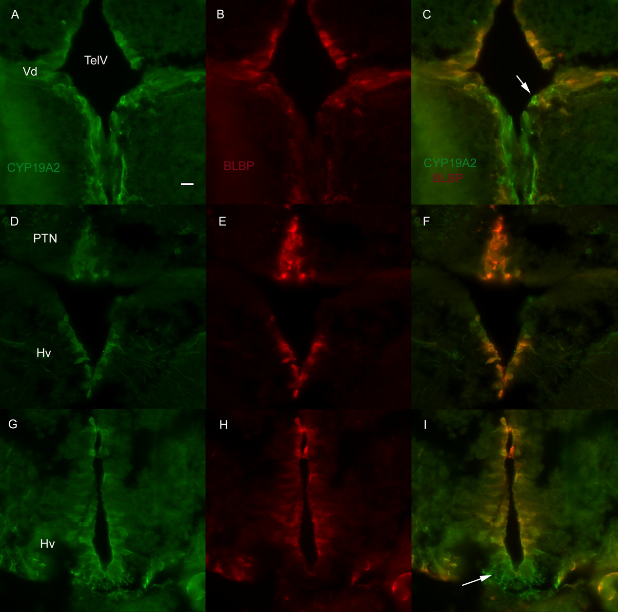
Fig. 8 Double immunofluorescence for CYP19A2 and BLBP. A-C: Most of the cells that surround the telencephalic ventricle (TelV) at the level of the dorsal nucleus (Vd) of the ventral telencephalic area that are positive for CYP19A2 are also positive for BLBP, with some exceptions (arrows). D-F: The majority of the cells in the posterior tuberal nucleus (PTN) and at least some levels of the ventral zone of the periventricular hypothalamus (Hv) are double positive. G-I: The part of the Hv that comes in contact with the pituitary is not double labeled (arrow in I). Scale bar = 10 μm.
Figure Data
Acknowledgments
This image is the copyrighted work of the attributed author or publisher, and
ZFIN has permission only to display this image to its users.
Additional permissions should be obtained from the applicable author or publisher of the image.
Full text @ Dev. Dyn.