
Fig. 2
- ID
- ZDB-IMAGE-071005-69
- Genes
- Publication
- Bateman et al., 2004 - Expression of the zebrafish Staufen gene in the embryo and adult
- All Figures
- Figures for Bateman et al., 2004
|
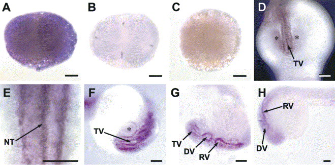
Fig. 2 Localization of staufen expression in the developing zebrafish embryo by whole mount in situ hybridization. (A–C) In situ hybridization was performed on 4-cell stage embryo (A). (B) Localization of vasa on 4-cell stage embryo. (C) A negative control in situ hybridization using the zebrafish staufen sense probe. In situ hybridization performed on 16 somite (D and E), 25 somite (F and G) and 24-h embryos (H). (D and E) The 16 somite embryo shows staufen staining in the telencephalic ventricle (TV) from the dorsal view. The neural tube (NT) is also staufen positive. (F and G) The 25 somite embryo shows staufen expression to be present in the telencephalic (TV), diencephalic (DV), rhombencephalic (RV) ventricles as well as in the neural tube. (H) The 24-h embryo continues to express staufen in the developing nervous system. The asterisk marks the eye vesicle. Magnification bar=100 μm.
Reprinted from Gene expression patterns : GEP, 5(2), Bateman, M.J., Cornell, R., d'Alencon, C., and Sandra, A., Expression of the zebrafish Staufen gene in the embryo and adult, 273-278, Copyright (2004) with permission from Elsevier. Full text @ Gene Expr. Patterns