
Fig. 4
- ID
- ZDB-IMAGE-071001-121
- Genes
- Publication
- Zecchin et al., 2007 - Distinct delta and jagged genes control sequential segregation of pancreatic cell types from precursor pools in zebrafish
- All Figures
- Figures for Zecchin et al., 2007
|
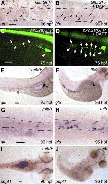
Fig. 4 Increase of enteroendocrine cell differentiation in mib mutants and DAPT-treated embryos. Panels A, B: in situ hybridization with a GFP antisense probe showing increased activity of the glucagon promoter in the gut (black arrows) of a glucagon:GFP (glu:GFP) transgenic zebrafish line treated with DAPT at 30 hpf (B). Panels C, D: nk2.2a:GFP transgenic embryo showing an increase of enteroendocrine cells (white arrows) when treated with DAPT at 30 hpf (D). The pancreatic duct (d) and the endocrine islet (arrowhead) are indicated. Panels E–H: glucagon expression in the gut (g) and pancreas (black arrowhead) of wt (E, G) and mib mutants (F, H) analyzed at 96 hpf. In panels G–H the gut is outlined. Panels I, J: expression of the peptide transporter pept1 in wt (I) and mib mutant embryos (J). Embryos are in lateral (C–F), ventral (A, B) or dorsal (G–J) views with anterior to the right. Scale bar is 50 μm.
Reprinted from Developmental Biology, 301(1), Zecchin, E., Filippi, A., Biemar, F., Tiso, N., Pauls, S., Ellertsdottir, E., Gnugge, L., Bortolussi, M., Driever, W., and Argenton, F., Distinct delta and jagged genes control sequential segregation of pancreatic cell types from precursor pools in zebrafish, 192-204, Copyright (2007) with permission from Elsevier. Full text @ Dev. Biol.