
Fig. 1
- ID
- ZDB-IMAGE-071001-118
- Genes
- Publication
- Zecchin et al., 2007 - Distinct delta and jagged genes control sequential segregation of pancreatic cell types from precursor pools in zebrafish
- All Figures
- Figures for Zecchin et al., 2007
|
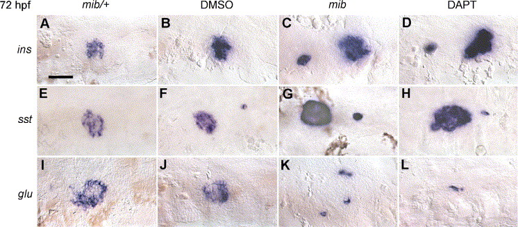
Fig. 1 Inhibition of Notch signalling increases differentiation of beta-cells. In situ hybridizations showing insulin (A–D) somatostatin (PPSS2) (E–H) and glucagon (I–L) expression in mib mutants and DAPT-treated embryos. The pancreatic area is depicted. Insulin and somatostatin expression, in blue, is increased in mib mutants and DAPT-treated embryos (C, D, G, H) compared with controls (A, B, E, F). Conversely, glucagon expression is reduced or absent (compare K, L with I, J). Embryos in panels A, E, I and B, F, J should be compared with embryos in panels C, G, K and D, H, L, respectively. Embryos have been hybridized at 72 hpf and are presented in a ventral view with anterior to the left. Scale bar in panel A is 50 μm.
Reprinted from Developmental Biology, 301(1), Zecchin, E., Filippi, A., Biemar, F., Tiso, N., Pauls, S., Ellertsdottir, E., Gnugge, L., Bortolussi, M., Driever, W., and Argenton, F., Distinct delta and jagged genes control sequential segregation of pancreatic cell types from precursor pools in zebrafish, 192-204, Copyright (2007) with permission from Elsevier. Full text @ Dev. Biol.