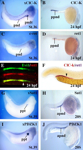
Fig. 3 Comparison of ClC-K, ret, Sat1, Evi1 and PDZK1 expression in Xenopus and zebrafish pronephros. The stages and probes analyzed are indicated in each figure. Anterior is toward the right. (A–D,G–J) Single in situ hybridization of the indicated markers. Note that in zebrafish ClC-K and ret1 expression is restricted to the posterior part of the duct while the proximal tubule specific markers Sat1 and PDZK1 are expressed in its anterior portion. (E) Comparison by fluorescent in situ hybridization of Evi1 (green) and ret1 (red) expression in a 24-hpf zebrafish embryo. Note that Evi1 expression extends further anteriorly than that of ret1. (F) Comparison by double in situ hybridization of ClC-K (red) and ret1 (purple) expression in a 24-hpf zebrafish embryo. Note that ClC-K expression extends further anteriorly than that of ret1. Abbreviations: apnd, anterior pronephric duct; pnd, pronephric duct; pndt, pronephric distal tubule; ppnd, posterior pronephric duct; ppt, proximal pronephric tubules.
Reprinted from Developmental Biology, 294(1), Van Campenhout, C., Nichane, M., Antoniou, A., Pendeville, H., Bronchain, O.J., Marine, J.C., Mazabraud, A., Voz, M.L., and Bellefroid, E.J., Evi1 is specifically expressed in the distal tubule and duct of the Xenopus pronephros and plays a role in its formation, 203-219, Copyright (2006) with permission from Elsevier. Full text @ Dev. Biol.

