
Fig. 13
- ID
- ZDB-IMAGE-070822-1
- Genes
- Publication
- Shi et al., 2006 - Zebrafish foxe3: Roles in ocular lens morphogenesis through interaction with pitx3
- All Figures
- Figures for Shi et al., 2006
|
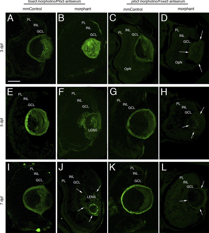
Fig. 13 Immunolocalization of Pitx3 and Foxe3 in foxe3 and pitx3 morphant eye tissues. The Pitx3 and Foxe3 proteins were immunolocalized in frozen sections of either foxe3 or pitx3 morphant eyes and the relevant mismatch control morpholino-injected embryos (mmControl). Pitx3 is detected in the lens of foxe3 mmControl embryos at 3, 5 and 7 dpf (panels A, E and I, respectively), and also in the foxe3 morphant lens at these same time points (panels B, F and J). The Pitx3 spatial expression pattern is disrupted in the foxe3 morphants. A wild-type Foxe3 expression pattern is displayed in the pitx3 mmControl embryos at 3, 5 and 7 dpf (panels C, G and K, respectively). In contrast, Foxe3 protein is not detected in the pitx3 morphants at 3, 5 and 7 dpf (panels D, H and L, respectively; arrows delineate the lens tissue). The scale bar in panel A (50 μm) applies to all the panels. Abbreviations: PL, photoreceptor layer; INL, inner nuclear layer; GCL, ganglion cell layer.
Reprinted from Mechanisms of Development, 123(10), Shi, X., Luo, Y., Howley, S., Dzialo, A., Foley, S., Hyde, D.R., and Vihtelic, T.S., Zebrafish foxe3: Roles in ocular lens morphogenesis through interaction with pitx3, 761-782, Copyright (2006) with permission from Elsevier. Full text @ Mech. Dev.