
IMAGE
Fig. 8
- ID
- ZDB-IMAGE-070802-34
- Genes
- Publication
- Kok et al., 2007 - The role of the SPT6 chromatin remodeling factor in zebrafish embryogenesis
- All Figures
- Figures for Kok et al., 2007
Image
|
Figure Caption
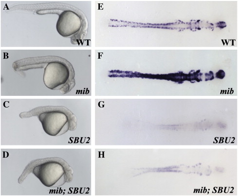
Fig. 8 mib;SBU2 double mutants reveal an epistatic interaction between Spt6 and Notch pathway. (A–D) Lateral views of 27-h-old embryos, anterior to the right. (E–H) Dorsal views of huC stained embryos, anterior to the right. All embryos were genotyped following photography. The phenotype of mib;SBU2 mutants (D) resembles SBU2 single mutants (C), rather than mib single mutant embryos (B). However, huC expression profile in mib;SBU2 double mutants (H) is stronger than SBU2 mutants (G) but not as strong as either wild-type (E) or mib single mutants (F).
Figure Data
Acknowledgments
This image is the copyrighted work of the attributed author or publisher, and
ZFIN has permission only to display this image to its users.
Additional permissions should be obtained from the applicable author or publisher of the image.
Reprinted from Developmental Biology, 307(2), Kok, F.O., Oster, E., Mentzer, L., Hsieh, J.C., Henry, C.A., and Sirotkin, H.I., The role of the SPT6 chromatin remodeling factor in zebrafish embryogenesis, 214-226, Copyright (2007) with permission from Elsevier. Full text @ Dev. Biol.