
Fig. S3
- ID
- ZDB-IMAGE-070620-32
- Genes
- Publication
- Hinits et al., 2007 - Mef2s are required for thick filament formation in nascent muscle fibres
- All Figures
- Figures for Hinits et al., 2007
|
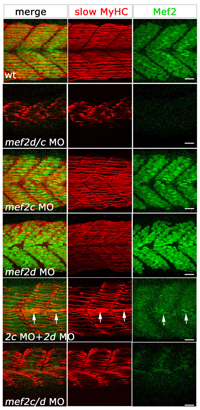
Fig. S3 Several morpholino combinations produce the same skeletal muscle phenotype. Confocal stacks of fluorescent immunodetection of slow MyHC (F59, red) and anti-Mef2 (green) antibodies of 24-hpf somites of control and embryos injected with different MO combinations, viewed in lateral flatmount. Phenotype severity in slow muscle fibres correlates with levels of Mef2 in fibre nuclei. Single morpholinos show no detectable defects in slow fibres and wild-type levels of Mef2 in nuclei. Double knockdown with either mef2d/c MO, mef2c+mef2d MOs or mef2c/d MO, shows a similar fibre phenotype. Note the correlation of residual Mef2 (arrows) with better MyHC accumulation. Scale bars: 20 μm.