
Fig. 2
- ID
- ZDB-IMAGE-070309-2
- Genes
- Publication
- Wilson et al., 2007 - Cadherin-4 plays a role in the development of zebrafish cranial ganglia and lateral line system
- All Figures
- Figures for Wilson et al., 2007
|
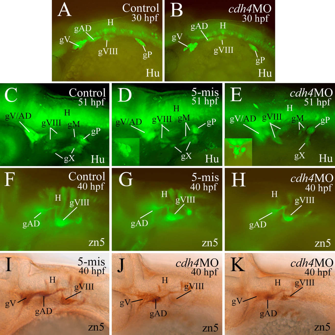
Fig. 2 Cranial and lateral line ganglia as revealed by anti-Hu immunostaining (Hu, A-E) and zn5 immunostaining (F-K). All panels are lateral views of whole mount zebrafish embryos with anterior to the left and dorsal up. A-H are from immunofluorescent methods, while panel I-K are from immunoperoxidase methods. The cdh4 morphant in H was from RcadMphB injections, while the remaining morphants were from RcadMphA injections. The trigeminal (gV) and anterodorsal lateral line ganglia (gAD) in D and E were out of focus, and their focused images are shown in their respective insets. J and K are from the same embryo with the former panel focusing on gV and gAD, while the latter one focuses on the statoacoustic ganglion (gVIII). gM, medial lateral line ganglion. Other abbreviations are the same as in Figure 1.