
Fig. 8
- ID
- ZDB-IMAGE-070307-63
- Genes
- Publication
- Hans et al., 2007 - Fgf-dependent otic induction requires competence provided by Foxi1 and Dlx3b
- All Figures
- Figures for Hans et al., 2007
|
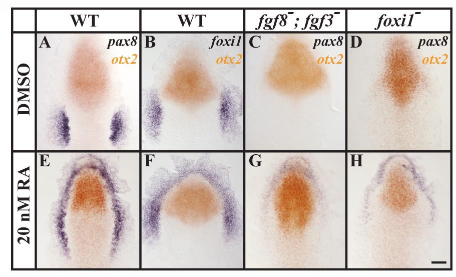
Fig. 8 Ectopic foxi1 expression after treatment with retinoic acid (RA) results in ectopic Fgf-dependent otic induction. (A, B, E, F) In comparison to wild-type control embryos treated with DMSO, embryos treated with 20nM RA show ectopic pax8 and foxi1 expression surrounding the anterior neural plate border without affecting the neural expression of otx2. (C, G) In fgf3, fgf8 double mutants, pax8 is completely abolished in the control embryos, whereas RA-treated double mutant embryos show weak anterior expression of pax8. (D, H) In foxi1 mutants treated with DMSO, pax8 expression can not be detected in the preotic region; but in foxi1 mutants treated with RA, residual anterior pax8 expression is present. Dorsal views of 1–3-somite stage embryos with anterior towards the top. Scale bar: 40 μm.