
|
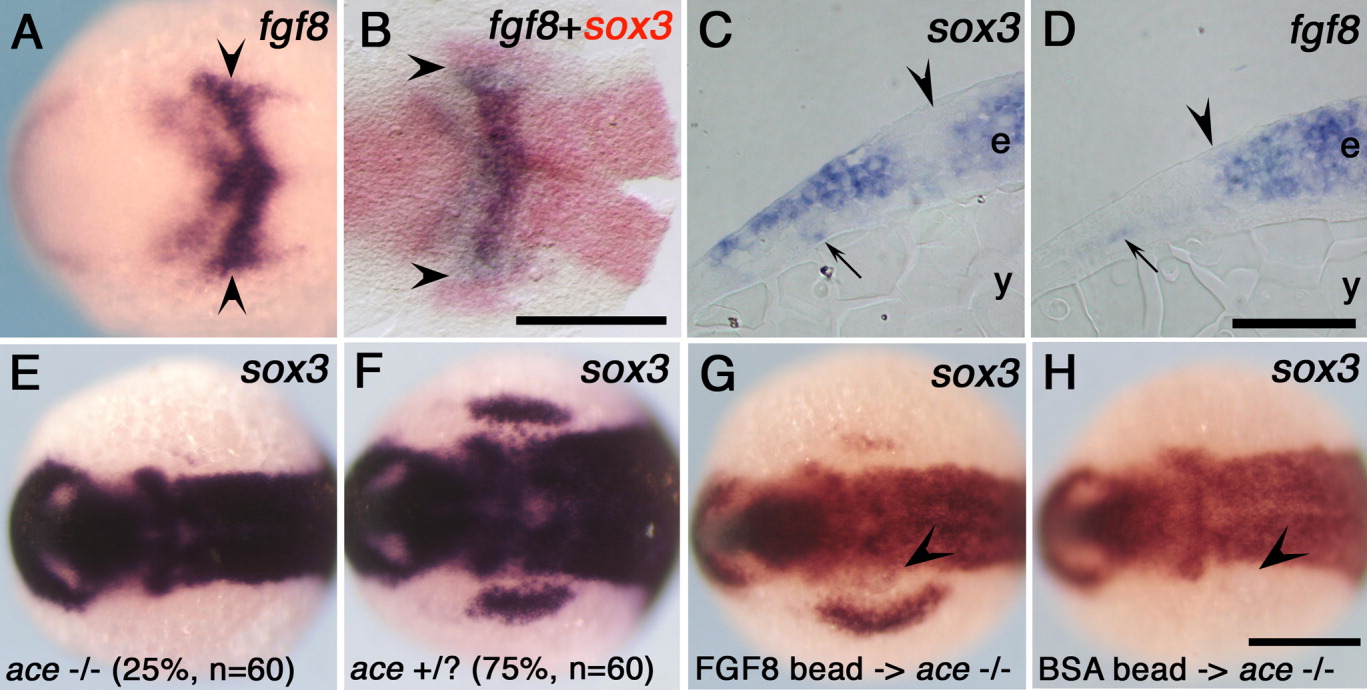
Fig. 2 Fibroblast growth factor-8 (FGF8) signal is required for development of the early placode. A: Expression of fgf8 in the anterior hindbrain region at the one-somite stage. B: Comparison of the expression of fgf8 (blue) and sox3 (red) at the one-somite stage. Arrowheads in A,B indicate the lateral border of the fgf8 domain in rhombomere 4 (r4). C,D: Transverse sections at the r4 level show intense expression of sox3 (C) and fgf8 (D) in the ectoderm at the one-somite stage. The edges of the neuroectoderm are marked by arrowheads. A few cells faintly expressing sox3 (C) or fgf8 (D) were observed in the mesendodermanl region (small arrows). E,F:sox3 expression in embryos obtained by intercross of ace heterozygous adults. Twenty-five percent of the obtained embryos (ace -/-) showed no placodal expression (E), while the others (75%, ace +/+ or ace +/-) showed normal sox3 expression (F). G,H:sox3 expression in the ace homozygous embryos implanted with FGF8b-soaked beads (G) and bovine serum albumin (BSA) -soaked beads (H). Arrowheads indicate the positions of the implanted beads. A,B,E-H: Dorsal views with anterior to the left. Genes examined are shown at the top right. e, ectoderm; y, yolk. Scale bars = 200 μm in A,B,E-H, 50 μm in C,D.