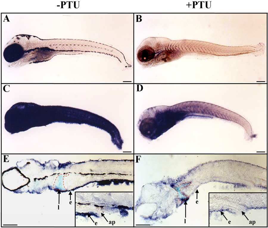
Fig. 1 Epidermal expression of rbp4 is down-regulated following treatment with PTU (1-phenyl-2-thio-urea). A-F: Embryos and larvae were raised, from fertilization to 10 days postfertilization (dpf), in water without (A,C,E) or with (B,D,F) added PTU to prevent pigment formation. A-F: Whole-mount in situ hybridizations are shown in lateral views and anterior to the left with digoxigenin-labeled sense (A,B) and antisense (C-F) rbp4 riboprobes. E,F: Parasagittal sections after whole-mount in situ hybridization. C-F: Epidermal rbp4 transcript hybridization signal was strongly reduced in the epidermis of PTU-treated larva (D,F) in comparison with untreated animals (C,E). E,F: The outline of the liver is traced in red. Inserts in E and F are enlarged views of the anal papilla region. e, epidermis; l, liver; ap, anal papilla. Scale bars = 250 μm.
Image
Figure Caption
Figure Data
Acknowledgments
This image is the copyrighted work of the attributed author or publisher, and
ZFIN has permission only to display this image to its users.
Additional permissions should be obtained from the applicable author or publisher of the image.
Full text @ Dev. Dyn.

