
|
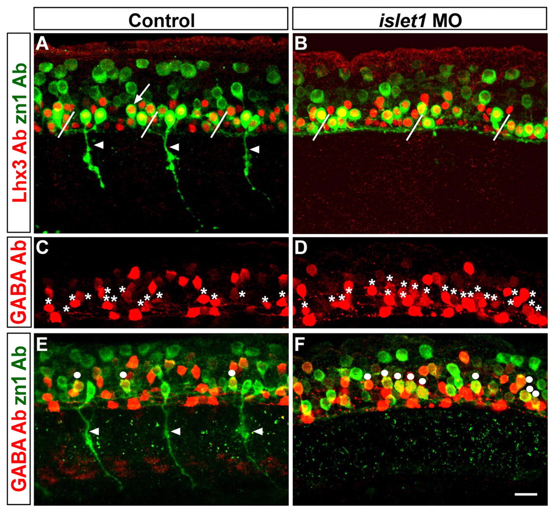
Fig. 4 Islet1 is required to inhibit interneuron formation. (A,B) Embryos co-stained with Lhx3 (red) and zn1 (green) Abs. (A) In control embryos, zn1 and Lhx3 were co-expressed in motoneurons that projected axons (arrowheads) out of the spinal cord and in VeLD interneurons (one is indicated by an arrow; slanted lines denote somite boundaries). (B) islet1 MO-injected embryos had cells that co-expressed Lhx3 and zn1, but they did not project axons out of the spinal cord, and instead had axons that projected caudally within the spinal cord. (C,D) Embryos stained with GABA Ab. (C) In control embryos, GABA Ab reveals KA'', KA', VeLD and other (unidentified) interneurons; cells in the V-K position are marked by asterisks. (D) Cells in the V-K position (asterisks) are more numerous in islet1 MO-injected embryos. (E,F) The same embryos shown in C and D, but here showing co-labeling with GABA (red) and zn1 (green) Abs. Dots indicate cells co-expressing GABA and zn1. (E) In control embryos, only a few cells co-express these markers. Arrowheads in E indicate CaP axons. (F) In islet1 MO-injected embryos there are many more cells that co-express zn1 and GABA. All embryos shown in this figure are at 28 hpf. Scale bar: 20 µm.