
Fig. 2
- ID
- ZDB-IMAGE-060605-29
- Genes
- Antibodies
- Publication
- Hutchinson et al., 2006 - Islet1 and Islet2 have equivalent abilities to promote motoneuron formation and to specify motoneuron subtype identity
- All Figures
- Figures for Hutchinson et al., 2006
|
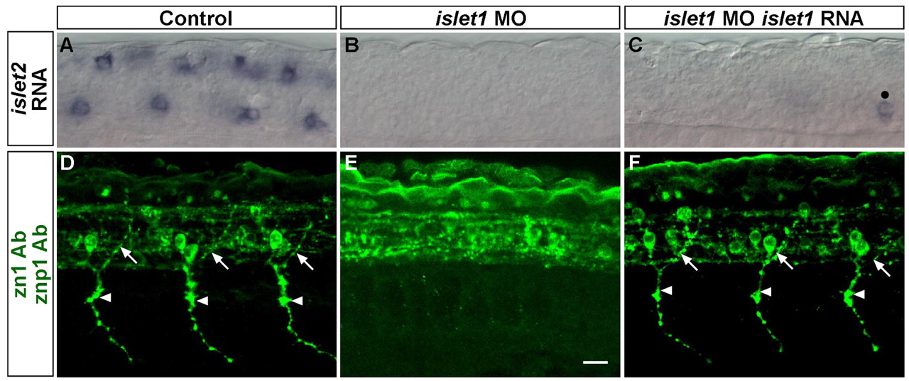
Fig. 2 Islet1 is required for PMN formation. (A-C) 20 hpf embryos stained with islet2 riboprobe. Control embryos (A) express islet2 in RBs (dorsally located cells) and CaPs (ventrally located cells). islet1 MO-injected embryos (B) lack islet2 expression. islet1 MO-injected embryos co-injected with islet1 RNA (C) also lack most islet2 expression; one islet2-positive PMN is indicated by a black dot. (D-F) 28 hpf embryos stained with zn1 and znp1 Abs (green). Control embryos (D) have dorsally projecting MiP axons (arrows) and ventrally projecting CaP axons (arrowheads). islet1 MO-injected embryos (E) lack both MiP and CaP axons. Co-injection of islet1 MO and islet1 RNA (F) restored both MiP and CaP axons. Scale bar: 20 µm.