
Fig. 1
- ID
- ZDB-IMAGE-051214-14
- Genes
- Publication
- Whitlock et al., 2005 - A role for foxd3 and sox10 in the differentiation of gonadotropin-releasing hormone (GnRH) cells in the zebrafish Danio rerio
- All Figures
- Figures for Whitlock et al., 2005
|
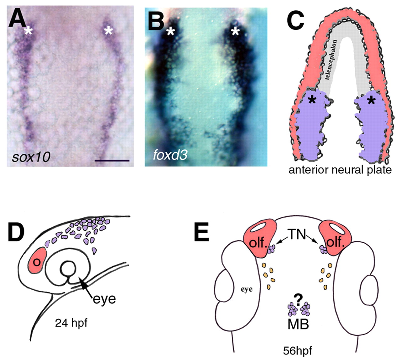
Fig. 1 Cranial neural crest domains in the developing zebrafish embryo. (A,B) Pattern of sox10 (A) and foxd3 (B) gene expression in 4- to 6-somite stage embryos (dorsal views). (C) The locations of the pre-migratory neural crest (purple) and placode (red) domains. Asterisks in A, B and C indicate the anterior limit of cranial neural crest domain. (D) Lateral view of a zebrafish head depicting the migratory routes of cranial neural crest (purple). Cranial neural crest cells migrating dorsal to the eye will contribute to the terminal nerve (TN) GnRH cells (E, purple) adjacent to the developing olfactory (olf) organ, and those migrating posterior to the eye may give rise to the midbrain (MB) GnRH cells (E, purple). Hypothalmic GnRH cells are indicated in orange (E). Scale bar in A: 100 µm for A,B.