
Image
|
Figure Caption
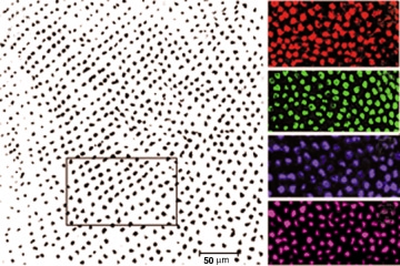
Fig. 4 Computational analysis of cone mosaic patterns. (A) Scion Image data file of an adult retinal whole mount processed for in situ hybridization (blue opsin), in which the area selected for analysis is boxed. (B-E) Confocal images of embryonic retinas at ~3dpf processed with red, green, blue or UV opsin probe (pseudo-colored), respectively.
Figure Data
Acknowledgments
This image is the copyrighted work of the attributed author or publisher, and
ZFIN has permission only to display this image to its users.
Additional permissions should be obtained from the applicable author or publisher of the image.
Full text @ Int. J. Dev. Biol.