
|
Image description by: Tanya Whitfield
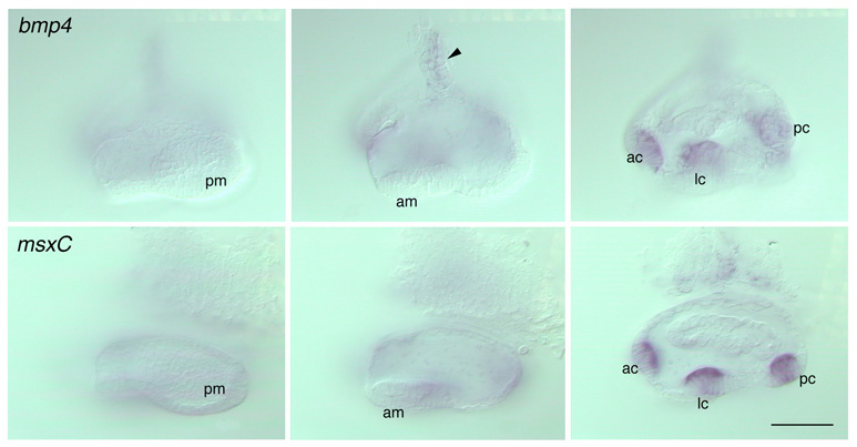
Anatomical structures shown: anterior and posterior maculae (unstained); anterior, lateral and posterior cristae (stained).
Stage: 60 h
Genetic (background) strain: none given
Genotype: phenotypically wild-type embryo from a dogtp85b/+ x dogtp85b/+ cross
Animal state: fixed
Labeling: bmp4 mRNA (top panels); msxC RNA (bottom panels)
Description: Expression of bmp4 (top panels) and msxC (bottom panels) in the zebrafish ear at 60h. Medial to lateral series (left to right panels) through dissected ears from whole mount preparations; DIC optics. The posterior macula (pm) is visible on the posteromedial wall of the ear. The anterior macula (am) is visible in cross section on the anteroventral floor of the vesicle; it appears as a thickened, pseudostratified region of epithelium. The upper row of nuclei belong to hair cells in this patch; the lower row of nuclei are those of the supporting cells. Neither macula expresses bmp4 or msxC at this stage. bmp4 stains a dorsal region of the otic vesicle at this stage (identity unknown), which sometimes appears duct-like, as in this preparation (arrowhead). The anterior, lateral and posterior cristae (ac, lc, pc) stain strongly for both bmp4 and msxC, and lie lateral to the two maculae.
Publication containing this image: T. Whitfield (unpublished).
| Preparation | Image Form | View | Direction |
| whole-mount | still | parasagittal | anterior to left |