
Fig. 6
- ID
- ZDB-FIG-260501-6
- Publication
- Szenker-Ravi et al., 2024 - CIROZ is dispensable in ancestral vertebrates but essential for left-right patterning in humans
- Other Figures
- All Figure Page
- Back to All Figure Page
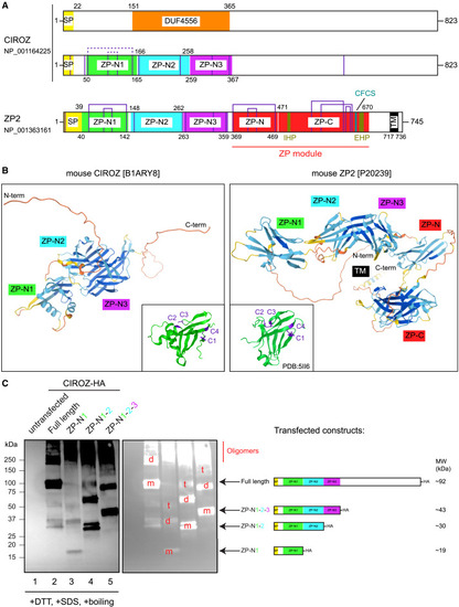
The N-terminal half of CIROZ bears homology to the 3 ZP-N domains found in ZP2 (A) Protein schematic structures of human CIROZ and human ZP2. The two proteins share the following domains: a signal peptide (SP; yellow), a zona pellucida N-terminal domain 1 (ZP-N1; green), ZP-N2 (cyan), and ZP-N3 (purple). ZP2 also contains a ZP module (ZP-N + ZP-C, red), internal and external hydrophobic patches (IHPs and EHPs, respectively; brown), a conserved furin cleavage site (CFCS; blue), and a transmembrane domain (TM; black). Purple vertical lines: conserved cysteines; known (full purple lines) and predicted (dashed purple lines) cysteine bonds are shown on top. (B) AlphaFold protein structure prediction for mouse CIROZ and mouse ZP2. The domains are indicated with the same color code as in (A). Insets: comparison of the ZP-N1 domain of CIROZ (AlphaFold2) to that of ZP2 (PDB: 5II6), highlighting the 4 conserved cysteines (purple). (C) Anti-HA Western blot analysis on conditioned media from HEK293T cells transfected with indicated human CIROZ-HA constructs. m, monomer; d, dimer; t, trimer; SDS, sodium dodecyl sulfate; DTT, dithiothreitol. |