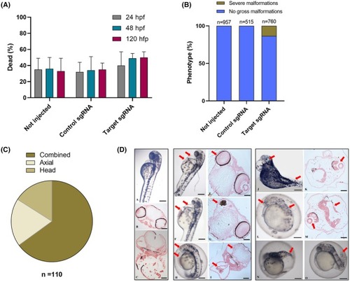
Mortality and phenotypic analysis of F0 embryos. (A) Embryo mortality rates were tracked at various hours postfertilization (hpf) for noninjected embryos and those microinjected with either scramble or target sgRNAs. The data, compiled from four independent microinjections, revealed variability in mortality across different clutches. However, the differences in mortality rates between the experimental groups did not reach statistical significance (mean ± SD, one‐way ANOVA). The total number of embryos analyzed was as follows: not injected, n = 1796; control sgRNA, n = 1030; and target sgRNAs, n = 1642. (B) Daily observation of embryos identified individuals with evident gross malformations, which were subsequently euthanized. This panel presents the cumulative percentage of embryos exhibiting severe phenotypes at 120 hpf, derived from three independent microinjections. The total number of embryos inspected for each group is indicated above the respective bars. (C) The severe malformations observed in embryos injected with target sgRNAs, as quantified in panel B (n = 110), are categorized into three main types: (a) head malformations, including instances of absent or duplicated heads and small or absent eyes. (b) Axial malformations: characterized by a lateral curvature of the body axis. (c) Miscellaneous: represents the largest category, encompassing embryos exhibiting multiple severe malformations occurring in combination. (D) Representative images illustrate severely malformed embryos (D to O) in comparison to control embryos (A–C). Edema accompanied by blood accumulation in the pericardial region (highlighted by the red arrows in panels D, F and H) is associated with abnormal eye (red arrows in panels E and G) and otic vesicle development and blood accumulation (red arrows in panel I). Severely impaired head and body development is highlighted by the red arrows in panels J and L (and corresponding sections in panels K and M). Abnormal axial development (red arrows) is observed in panels N and O. Scale bars are 100 μm for whole mounts and 50 μm for sections.
|
