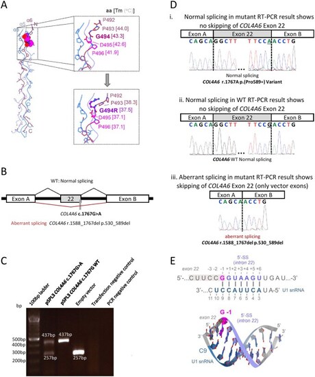
Variant-specific characterization: protein structure modeling and minigene assay of the COL4A6 c.1767G>A variant. (A) Structural consequences of the Gly494Arg substitution. (B) The COL4A6 exon 22 trapping vector construct for the in vitro splice assay of c.1767G>A variant with a schematic summary of results. An amplicon containing either mutated or wild-type (WT) exon 22 was inserted between exons A and B in the pSPL3 vector. The black lines above the figure show the wild-type results, while the red lines below summarize the aberrant splicing results. (C) Gel electrophoresis of cDNA amplicons from HEK293T cells transfected with the c.1767G>A variant-containing vector, WT, or empty vector. The WT and mutant cDNA amplicons revealed correctly spliced 437 bp amplicons (with less intensity in mutant sample). The mutant also yielded a 257 bp amplicon, identical to the empty vector. Controls performed as expected. (D) A schematic of the resulting splice product sequences. The WT and mutant amplicons produce a 437 bp normally spliced product (i, ii) variant position highlighted in the yellow box. The mutant amplicon additionally shows the skipping of exon 22 (iii), indicated by the sequence of the vector exons A and B. (E) Proposed mechanism of the aberrant splicing caused by the c.1767G>A variant. Our modeling indicates that the substituted nucleobase (magenta, G-1) maps to the exon 22-intron 22 junction of COL4A6 and base pairs with C9 of the U1 snRNA, thereby contributing to the formation of the U1-5′ splice site (SS) duplex. The structural model of the U1-5′ SS duplex (bottom panel), formed at the exon 22-intron 22 junction of COL4A6, is shown in stick representation. The base-paired pre-mRNA and U1 nucleobases are colored light blue and teal, respectively. Abbreviation: WT, wild-type.
|
