
Fig. 4
- ID
- ZDB-FIG-260401-130
- Publication
- Dereddi et al., 2026 - Oligodendrocyte mechanotransduction channel TMEM63A regulates myelin sheath geometry
- Other Figures
- All Figure Page
- Back to All Figure Page
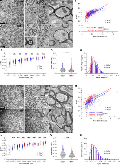
TMEM63A is required for fine-tuning radial myelin sheath geometry (A–D) Electron micrographs of CC from T63a+/+ (A) and T63a−/− (C) mice at P35, with higher magnifications of boxed areas to the right. Insets, unmyelinated T63a+/+ and myelinated T63a−/− small-diameter axons (<0.2 μm; white outline). Note the normally myelinated T63a+/+ (B) vs. hypomyelinated T63a−/− large-diameter axons (D). Dashed lines demarcate myelin area. (E and F) g ratios of individual myelinated axons as a function of axon diameter (E) and binned axon diameter (F, in μm) in the CC at P35. Slopes, p < 0.0001 (E). Boxed area (F) indicates the absence of 0.1–0.2 μm myelinated axons in controls. (G and H) Myelin area of individual myelinated axons (G, in μm2) and relative frequency distribution (with Gaussian fit, H) in the CC at P35. (I–L) Electron micrographs of CC from T63a+/+ (I) and T63a−/− (K) mice at P60, with higher magnifications of boxed areas to the right. Insets, unmyelinated T63a+/+ and myelinated T63a−/− small-diameter axons (<0.15 μm; white outline). Note the normally myelinated T63a+/+ (J) vs. hypomyelinated T63a−/− large-diameter axons (L). Dashed lines demarcate myelin area. (M and N) g ratios of individual myelinated axons as a function of axon diameter (M) and binned axon diameter (N, in μm) in the CC at P60. Slopes, p = 0.3025; intercepts, p < 0.0001 (M). Boxed area (N) indicates the absence of 0.05–0.15 μm myelinated axons in controls. (O and P) Myelin area of individual myelinated axons (O, in μm2) and relative frequency distribution (with Gaussian fit, P) in the CC at P60. Data represent mean ± SD (F and N) and median with interquartile range (G and O). ∗∗p < 0.01, ∗∗∗∗p < 0.0001 (including H and P). See also Figure S4 and Table S3 (sample sizes and statistical tests). |