FIGURE

Figure 5

ID
ZDB-FIG-260313-16
Publication
Tarahomi et al., 2025 - Transcriptomic identification and developmental mapping of nrg3b expression in zebrafish
Other Figures
All Figure Page
Back to All Figure Page
Figure 5

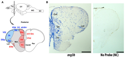
Spatial expression of nrg3b in the adult zebrafish telencephalon. A, The schematic view of the adult zebrafish forebrain shows the telencephalon’s major subregions in a coronal section. Areas corresponding to the dorsal (Dd, Dm, Dp) and ventral (Vd, Vv) domains of the telencephalon are indicated. Adapted conceptually from the telencephalic atlas published by (Diotel et al., 2015). B, ISH on coronal brain sections revealed expression of the nrg3b gene predominantly in the dorsomedial (Dm), ventral (Vd), and ventro-ventral (Vv) nuclei of the telencephalon. Scale bars: 50 μm. Stained tracts and nuclei overlapped with regions composed of rich populations of neurons and glial cells, which are involved in adult neurogenesis and the organization of neural circuits. The Lot was visible and served as an anatomical reference. No staining was observed in negative-control sections processed without probe (NC), confirming the specificity of the signal.

Expression Data
Gene:
Fish:
Anatomical Terms:
Stage: Adult

Expression Detail
Antibody Labeling
Phenotype Data

Phenotype Detail
Acknowledgments
This image is the copyrighted work of the attributed author or publisher, and ZFIN has permission only to display this image to its users. Additional permissions should be obtained from the applicable author or publisher of the image. Full text @ Turk J Biol