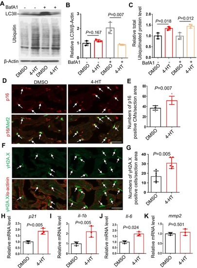
Cardiomyocyte‐specific overexpression of fabp7a gene caused impaired protein homeostasis and accelerated cellular senescence. (A–C) Western blot (A) and quantification analysis of the LC3 II (B) and ubiquitinated proteins (B) levels indicated fish heart treated with or without 50 nM BafA1 for 4 h. n = 3 biological replicates, one‐way ANOVA. (D, E) Representative images of immunofluorescence using anti‐p16 antibody co‐stained with anti‐Mef2 antibody (D) and quantification of the numbers of anti‐p16/anti‐Mef2 signal (E) in the cardiomyocyte‐specific overexpression of Fabp7a fish at 3 months post‐4‐HT induction. Arrows point to overlapping signals. Scale bars: 20 μm. n = 5, one‐way ANOVA. (F, G) Representative images of immunofluorescence using anti‐γH2A.X antibody co‐stained with anti‐α‐actinin antibody (F) and quantification of the numbers of anti‐γH2A.X/anti‐α‐actinin co‐stained cell signal (G) in the cardiomyocyte‐specific overexpression of Fabp7a fish at 3 months post‐4‐HT induction. Scale bars: 20 μm. n = 5, one‐way ANOVA. (H–K) Quantitative RT‐PCR analysis of cellular senescence marker p21 and SASP markers including il‐1b, il‐6, and mmp2 in the cardiomyocyte‐specific overexpression of Fabp7a fish at 3 months post‐4‐HT induction. n = 3 biological replicates, one‐way ANOVA.
|
