FIGURE

Figure 1

ID
ZDB-FIG-251028-39
Publication
Määttä et al., 2025 - Utilizing CRISPR-Cas13d-knockdown in zebrafish to study a rare monogenic bone fragility syndrome
Other Figures
All Figure Page
Back to All Figure Page
Figure 1

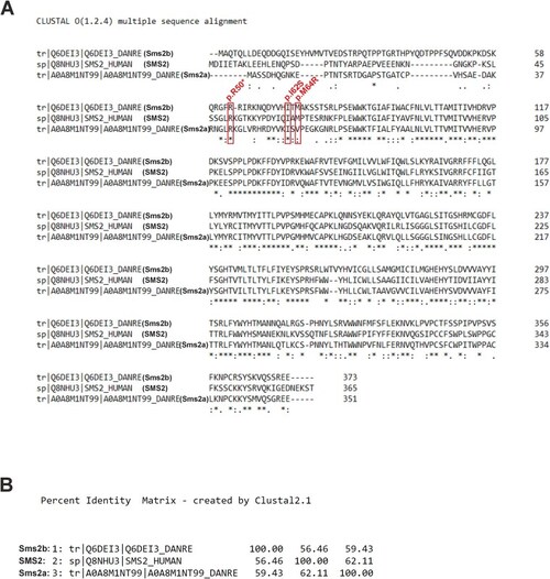
Protein sequence comparison of SMS2, Sms2a, and Sms2b. (A) Multiple sequence alignment of human SMS2 (365 aa) and zebrafish Sms2a (351 aa) and Sms2b (373 aa). Residues are identical at 165 positions (marked as stars). Positions of three human SMS2 pathogenic variants (p.R50*, p.I62S, p.M64R)4 have been indicated with boxes. Human SMS2 residues R50 and I62 are detected in both Sms2a and Sms2b sequences. Human SMS2 residue M64 is detected only in Sms2b sequence. “*” means that the residues in that column are identical in all sequences in the alignment; “:” means that conserved substitutions have been observed, that is, amino acid is replaced by one having similar characteristics; “.” means that semi-conserved substitutions are observed, that is, amino acids having similar shape. (B) Protein sequence percent identity matrix. Sms2a and Sms2b share 59% sequence identity. Sms2a and SMS2 share 62% sequence identity. Sms2b and SMS2 share 56% sequence identity. The following UniProt sequences were used: SMS2, Q8NHU3; Sms2a, A0A8M1NT99; Sms2b, Q6DEI3. The protein sequences were compared in the UniProt Knowledgebase35 using the Clustal Omega for multiple sequence alignment36 and Clustal 2.1 for percent identity matrix.

Expression Data

Expression Detail
Antibody Labeling
Phenotype Data

Phenotype Detail
Acknowledgments
This image is the copyrighted work of the attributed author or publisher, and ZFIN has permission only to display this image to its users. Additional permissions should be obtained from the applicable author or publisher of the image. Full text @ JBMR Plus