
Fig. 5
- ID
- ZDB-FIG-251016-90
- Publication
- Park et al., 2025 - Lego Self-Assembly Model of Zebrafish RGB Cone Pentamer Formation: Unique Role of Crumbs2b in Cone Coalescence via Planar Orientational Cell Adhesions
- Other Figures
- All Figure Page
- Back to All Figure Page
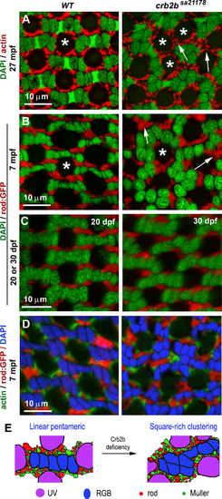
Crb2b deficiency results in the disorganization of rods, UV cones, and Müller glial cells, as revealed by transverse imaging of the dorsal intermediate retina. (A) Actin staining (red) revealed that the apical processes of Müller glial cells surround UV cones regularly (asterisks) in the wild-type but often form larger clusters in crb2bsa21178 homozygous mutants. (B, C) Rods, revealed by GFP expression (red) in the Tg(-3.7rho:EGFP)kj2 transgenic background,50 formed linear clusters separating individual pentamers in the wild-type. However, they formed larger irregular clusters in the mutants (arrows) at 7 mpf (B) but not at 1 mpf (C). (D) Simultaneous visualization of actin, rods, and nuclei (DAPI staining) revealed that pentameric cone units were surrounded by rods and Müller glial cell processes. (E) Diagrams summarize the differences in the patterning of all photoreceptor types between the wild-type and crb2bsa21178 homozygous mutant retinas (n = 4 fish for each condition). |