
Fig. 1
- ID
- ZDB-FIG-251006-48
- Publication
- Li et al., 2025 - EMB is essential for enteric nervous system development mediated by PI3K signaling
- Other Figures
- All Figure Page
- Back to All Figure Page
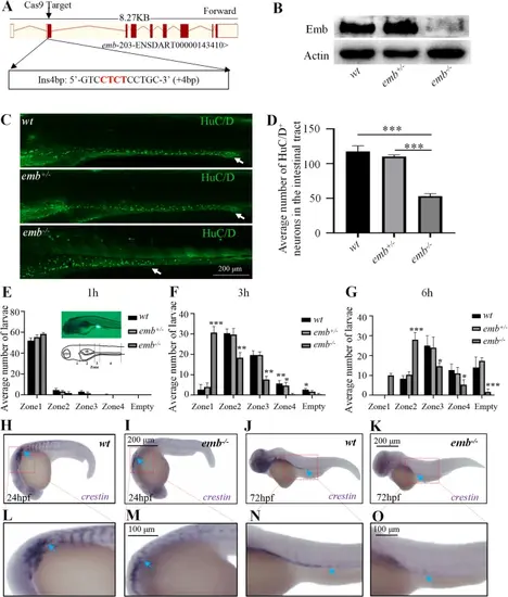
Loss of emb causes ENS defects in zebrafish. A, Generation of targeted emb mutant in Zebrafish. A 4 bp insertion in Exon 2 was introduced through the CRISPR/Cas9 system. B, Expression of the Emb protein is disrupted in different genotypes determined by western blots. C, The enteric neurons at 5 dpf zebrafish larva of wild type (wt), heterozygous and homozygous emb mutants are labeled by HuC/D and white arrows point to the most posterior margin where the enteric neurons migrated to. D, Quantification of the numbers of HuC/D positive cells in the intestinal tract, ***, p < 0.001, one-way ANOVA with Bonferroni post-hoc, N = 10 for each group. E-G, Quantification of the number of larvae in each group based on the anterior extent of tracer. “empty” means fluorescent tracers are completely evacuated in the intestine. Statistical significances compared to wt are labeled on top of each bar, *, p < 0.05; **, p < 0.01; ***, p < 0.001, one-way ANOVA with Bonferroni post-hoc, N = 60. H-K, Lateral view of in situ hybridization results of 24 hpf and 72 hpf larvae using crestin probe, blue arrows point to migrating neural crest cells. L-O, Locally enlarged images of the H–K images (Red dashed box) |