
Fig. 5
- ID
- ZDB-FIG-250926-26
- Publication
- Rutter et al., 2025 - Retinopathy-associated inosine monophosphate dehydrogenase 1 mutations cause metabolic and filament defects in cones
- Other Figures
- All Figure Page
- Back to All Figure Page
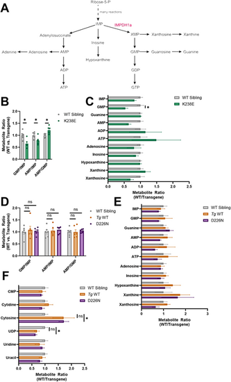
Purine or pyrimidine steady-state metabolite levels are affected in zebrafish with Impdh1a mutations. (A) Schematic showing de novo purine biosynthesis. IMPDH1a is highlighted in red. (B) Steady-state metabolite ratios in K238E mutant zebrafish compared to WT sibling eyes. GMP/IMP and AMP/IMP were lower in K238E mutant zebrafish compared to WT siblings (P=0.015 and P=0.027, respectively), whereas AMP/GMP was higher in K238E mutants (P=0.027). n=6 for both groups. Significance determined by unpaired two-tailed t-tests with Holm-Šídák correction for multiple comparisons. Error bars are s.e.m. (C) Ratio of purine-related metabolites for K238E mutant or WT sibling zebrafish eyes. GMP was significantly lower in K238E mutant zebrafish compared to WT siblings. n=6 for both groups. *P=0.048 (unpaired two-tailed t-tests with Holm-Šídák correction for multiple comparisons). Error bars are s.e.m. (D) Steady-state metabolite ratios in D226N mutant or Tg WT compared to WT sibling zebrafish retinas. Ratios in D226N mutant and Tg WT zebrafish retinas were not significantly different from those in WT sibling retinas. n=6 for all groups. ns, not significant (unpaired two-tailed t-tests with Holm-Šídák correction for multiple comparisons). Error bars are s.e.m. (E) Ratios of purine-related metabolites in D226N mutant, Tg WT or WT sibling zebrafish retinas. There were no significant differences in purine metabolites between mutant and WT retinas (unpaired two-tailed t-tests with Holm-Šídák correction for multiple comparisons). n=6 for all groups. Error bars are s.e.m. (F) Ratios of pyrimidine-related metabolites in D226N mutant, Tg WT or WT sibling zebrafish retinas. In retinas from D226N mutant zebrafish, cytosine was significantly higher (P=0.046), and UDP was significantly lower (P=0.049), than in WT sibling retinas, but there were no differences in cytosine (P=0.49) and UDP (P=0.26) between Tg WT and WT sibling retinas. n=6 for all groups. Significance determined by unpaired two-tailed t-tests with Holm-Šídák correction for multiple comparisons. Error bars are s.e.m. |