
Fig. 4
- ID
- ZDB-FIG-250903-64
- Publication
- Lange et al., 2025 - Genetic compensation highlights the importance of neural cell adhesion molecule Ncam1 paralogs in balancing signaling pathways during zebrafish lateral line development
- Other Figures
- All Figure Page
- Back to All Figure Page
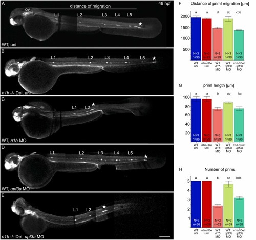
Knockdown of upf3a in ncam1b mutants partially phenocopies the ncam1b knockdown phenotype. (A–E) Lateral views of 48 hpf Tg(ClaudinB::lynGFP) (A) Wild type uninjected (WT uni) (B) n1b -/- Del uninjected, (C) n1b MO in WT, (D) upf3a MO in WT and (E) upf3a MO in n1b -/- Del. Embryos are oriented with rostral to the left and dorsal to the top. (A) At 48 hpf, primI (star) of WT reaches the tip of the tail and deposits 5 proneuromasts (L1–L5). The migration distance was measured from the otic vesicle (ov) to primI. (B) n1b -/- Del mutants show the same phenotype as WT, with primI reaching the tip of the tail and 5 deposited proneuromasts. (C) ncam1b MO injection leads to reduced migration of a smaller primI and deposition of fewer proneuromasts. (D) upf3a MO injection in WT does not induce any defects in lateral line system development. (E) upf3a MO injection in n1b -/- Del leads to reduced primI migration and fewer deposited neuromasts. (F–H) Quantification of migration distance, primI length and number of proneuromasts. Bars indicate mean values of relative numbers, with error bars representing standard error. Differences between the five groups were analyzed using a one-way ANOVA followed by Scheffé post hoc tests. Differences were considered significant if the F-value exceeded the critical Scheffé value (p < 0.05). Groups that share the same letter do not differ significantly. Groups with different letters show significant differences in their means. Scale bar 200 µm. Abbreviations: WT: Wild type; n1b -/- Del: ncam1b deletion mutant; MO: Morpholino; uni: uninjected; primI: primordium; pnm: proneuromast, ov: otic vesicle. |