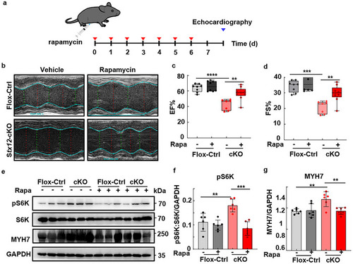
Rapamycin treatment relieved cardiac failure in Stx12‐cKO mice. a) Schematic diagram of rapamycin injection in mice. Cardiac functions were evaluated by echocardiography after intraperitoneally daily injection in Stx12‐cKO (Stx12flox/flox with CTnT‐Cre) and control (Stx12flox/flox) mice for one week. b) Representative M‐mode echocardiography of left ventricular chamber of control (Stx12‐flox) and Stx12‐cKO mice after rapamycin or vehicle treatment. c,d) Changes in ejection fraction (EF%) (c) and fractional shortening (FS%) (d) of the left ventricle in control (Stx12‐flox) and Stx12‐cKO mice after rapamycin or vehicle treatment. EF% was significantly increased in Stx12‐cKO mice after rapamycin treatment (Flox‐control + vehicle, n = 8; Flox‐control + rapamycin, n = 8; cKO + vehicle, n = 7, t‐test, p = 0.0003; cKO + rapamycin, n = 9, p = 0.0025). FS% was significantly increased in Stx12‐cKO mice after rapamycin treatment (Flox‐control + vehicle, n = 8; Flox‐control + rapamycin, n = 8; cKO + vehicle, n = 7, p = 0.0003; cKO + rapamycin, n = 9, p = 0.0025). e) Western blot analysis of S6K, phosphorylated S6K, and MYH7 in control (Stx12‐flox) and Stx12‐cKO mice, with GAPDH as the loading control. f) Quantification of Western blot of phosphorylated S6K (Flox‐control + vehicle, n = 6; Flox‐control + rapamycin, n = 6; cKO + vehicle, n = 6; cKO + rapamycin, n = 6; Flox‐control + vehicle vs cKO + vehicle, p = 0.00373; cKO + vehicle vs cKO + rapamycin, p = 0.0020). g) Quantification of Western blot of MYH7 (Flox‐control + vehicle, n = 6; Flox‐control + rapamycin, n = 6; cKO + vehicle, n = 6; cKO + rapamycin, n = 6; Flox‐control + vehicle vs cKO + vehicle, p = 0.0037; cKO + vehicle vs cKO + rapamycin, p = 0.0057). Statistical results: **p < 0.01, ***p < 0.001, ****p < 0.0001; t‐test.
|
