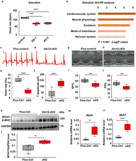
STX12 deficiency caused heart failure in zebrafish and mice. a) Quantification of the heart rates of zebrafish embryos. Heart rate was significantly decreased after Stx12 knockdown (KD) in zebrafish (control, n = 10, KD‐1, n = 10, t‐test, p < 0.0001; KD‐2, n = 10, p < 0.0001). bpm, beats per minute. b) GO‐HP analysis of zebrafish RNAseq data suggested abnormalities of the cardiovascular system after Stx12 knockdown. c,d) Electrocardiograms (ECG) of control (Stx12‐flox) (c) and cardiac‐specific Stx12 knockout mice (d). Scale bars: horizontal 20 ms, vertical 100 µV. e) Quantification of heart rate derived from ECG. Heart rate in Stx12 knockout mice was significantly decreased compared to wild type (Flox‐control, n = 5, CKO, n = 6, p < 0.0001). f) Comparison of T wave amplitude. T wave amplitude was significantly increased in Stx12‐cKO (Stx12flox/flox with CTnT‐Cre) mouse electrocardiograms compared to control (Stx12flox/flox) (Flox‐control, n = 6, cKO, n = 6, p < 0.0001). g,h) Representative M‐mode echocardiography of left ventricular chamber in control (Stx12‐flox) (g) and Stx12 cardiac‐specific knockout mice (h). i,j) Measurement of ejection fraction (EF%) (i) and fractional shortening (FS%) (j) of Stx12 cKO mice and control wild‐type mice. Left ventricular EF% in Stx12‐CKO mice was significantly decreased (Flox‐control, n = 9, cKO, n = 8, p = 0.0006). Left ventricular FS% in Stx12‐CKO mice significantly decreased (Flox‐control, n = 9, cKO, n = 8, p = 0.0004), indicating a decrease in ventricular contractile function. k) Western blot analysis of endogenous MYH7/β‐MHC level in control (Stx12‐flox) and Stx12‐cKO mice hearts. GAPDH was used as a loading control. l) Quantification of Western blot in (k). Myh7 level was significantly decreased compared with the wild type (Flox‐control, n = 7, cKO, n = 5, p = 0.0015). m,n) qRT‐PCR analysis of cardiac Nppb (m) and Myh7 (n) mRNA levels in control (Stx12‐flox) and Stx12‐cKO mice heart (n = 6 mice per group). There were significant increases of the mRNA level of Nppb and Myh7 in Stx12‐cKO mice (Nppb: Flox‐control, n = 6, cKO, n = 6, p = 0.0011; Myh7: Flox‐control, n = 6, cKO, n = 6, p < 0.0001), indicating myocardial hypertrophy and heart failure. Statistical results: **p < 0.01, ***p < 0.001, ****p < 0.0001; t‐test.
|
