
Fig. 3
- ID
- ZDB-FIG-250825-94
- Publication
- Wang et al., 2025 - Cytoneme-mediated intercellular signaling in keratinocytes is essential for epidermal remodeling in zebrafish
- Other Figures
- All Figure Page
- Back to All Figure Page
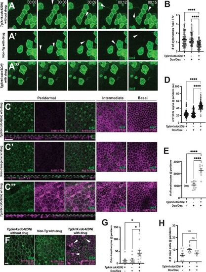
Cytoneme-mediated signaling regulates keratinocyte differentiation and proliferation. (A–A”) Still images from time-lapse movies demonstrate that cytoneme extension (arrowheads) is inhibited in keratinocytes expressing cdc42DN. (B) Expression of cdc42DN effectively inhibits cytoneme extension (F2, 231 = 25.38, p<0.0001, N=234 cells, 10 larvae total). (C) Periderm layer of Tg(krt4:TetGBDTRE-v2a-cdc42DN;krt4:lyn-EGFP;krtt1c19e:lyn-tdTomato) without drug treatment. (C’) Periderm layer of Tg(krt4:lyn-EGFP;krtt1c19e:lyn-tdTomato) with drug treatment. (C’’) Periderm layer of Tg(krt4:TetGBDTRE-v2a-cdc42DN;krt4:lyn-EGFP;krtt1c19e:lyn-tdTomato) with drug treatment exhibits disorganization. Intermediate keratinocytes were not depleted, and basal stem cells were unaffected by the manipulation. Note that, unlike the other two controls, the epidermis was not flat, resulting in partial display of basal cells in the intermediate layer and dark areas in the basal layer. Cross-section view exhibits krtt1c19e expression at the periderm. (D) Cytoneme inhibition increases krtt1c19e expression in the periderm (F2, 387 = 226.7, p<0.0001, N=39 larvae total) and leads to an increased number of keratinocytes within the periderm (E) (F2, 18 = 27.36, p<0.0001, N=21 larvae total). (F–G) Significant increase in proliferating peridermal keratinocytes in cdc42DN expressing animals (F2, 28 = 4.888, p=0.0151, N=9 larvae). (H) The cell death rate in the periderm is not affected by cytoneme inhibition, tested with acridine orange incorporation (F2, 17 = 3.192, p=0.0666, N=20 larvae total). Statistical significances were assessed by One-way ANOVA followed by Tukey’s HSD post hoc test. Scale bars: 20 µm (A–A”, C–C”, F). Error bars indicate mean ± SEM. |