
Fig. 2
- ID
- ZDB-FIG-250825-93
- Publication
- Wang et al., 2025 - Cytoneme-mediated intercellular signaling in keratinocytes is essential for epidermal remodeling in zebrafish
- Other Figures
- All Figure Page
- Back to All Figure Page
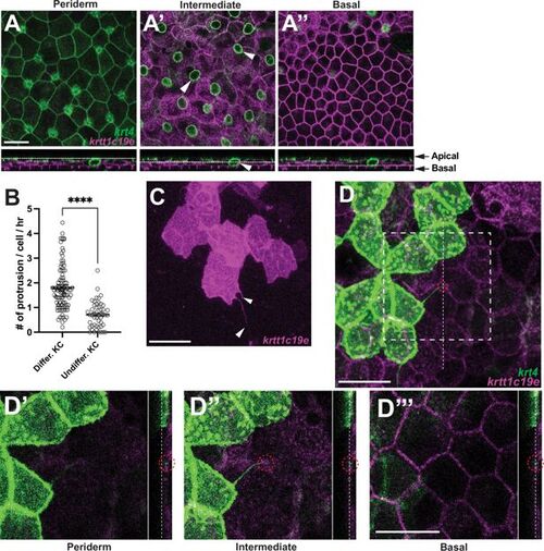
Keratinocyte cytonemes establish physical contact with underlying undifferentiated keratinocytes. (A–A”) Postembryonic zebrafish epidermis of Tg(krt4:lyn-EGFP;krtt1c19e:lyn-tdTomato) comprises three distinct layers. (A) Expression of lyn-EGFP in krt4+ cells in the periderm layer. (A’–A”) Expression of lyn-tdTomato (pseudo-colored magenta) in krtt1c19e+ cells in (A’) the intermediate layer and (A”) the basal layer. Note that the green circular labeling in (A’, arrowheads) represents mucous-secreting goblet cells. Chang and Hwang, 2011 (B, C) Undifferentiated keratinocytes (KC) extend significantly fewer cytoneme-like protrusions (arrowheads, p<0.0001, N=49, 3 larvae; N=91 cells, 6 larvae total). (D–D”’) Cytonemes from fully differentiated keratinocytes make physical contact with undifferentiated keratinocytes in the intermediate layer (red dotted circles). Note that dotted lines indicate the layers in the cross-sectional views. Statistical significances were assessed by Student t test. Scale bars: 20 µm (A–A”, C, D–D”’). Error bars indicate mean ± SEM. |