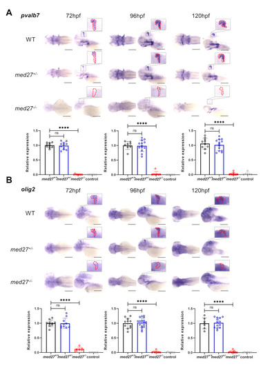
med27 LoF resulted in severe cerebellar atrophy. The results presented here are from the INS10 mutant line. For results from the INS5 and DEL5 mutant lines, refer to Fig. S5 and S6. Representative dorsal and lateral views of WISH for pvalb7 (A) and olig2 (B) at 72 hpf (pavlb7: med27+/+n = 10, med2+/−n = 11, med27−/−n = 9, control n = 6; olig2: med27+/+n = 10, med27+/−n = 10, med27−/−n = 6, control n = 6), 96 hpf (pavlb7: med27+/+n = 8, med27+/−n = 13, med27−/−n = 7, control n = 6; olig2: med27+/+n = 11, med27+/−n = 17, med27−/−n = 7, control n = 6), and 120 hpf (pavlb7: med27+/+n = 10, med27+/−n = 13, med27−/−n = 13, control n = 6; olig2: med27+/+n = 7, med27+/−n = 13, med27−/−n = 9, control n = 6) in larvae of the three genotypes. Enlarged views highlight the cerebellar region on lateral views enclosed by dashed red lines, which were quantified for gene expression comparison. Error bars represent mean ± SD. Statistical analysis was performed using one-way ANOVA. ns: not significant; ****: P<0.0001
|
