
Fig. 5
- ID
- ZDB-FIG-250508-20
- Publication
- Zhao et al., 2025 - Peptide-Modified Lipid Nanoparticles Boost the Antitumor Efficacy of RNA Therapeutics
- Other Figures
- All Figure Page
- Back to All Figure Page
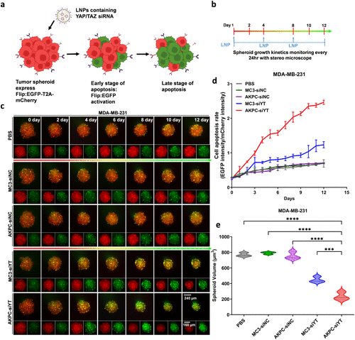
Antitumor effect of AKPC-siYT in 3D tumor spheroids of MDA-MB-231. a, Schematic representation of the plasmid containing EGFP-T2A-Caspase3-mCherry (cell apoptosis sensor). b, Schematic representation of seeding and treatment of spheroids to monitor spheroid growth kinetics. On days 1, 4, and 8, LNPs were used to treat MDA-MB-231-derived spheroids in different groups (siRNA, 2 μg/mL). A stereo microscope was used to record the tumor spheroids each day for 12 consecutive days. c, Images of representative MDA-MB-231 spheroids over time after treatments with LNPs (MC3-siNC/AKPC-siNC: LNPs contain a negative control of siRNA; MC3-siYT/AKPC-siYT: LNPs contain a siYAP and siTAZ mixture), Flip:EGFP intensity (in green) represents cell apoptosis. d, Kinetics of the cell apoptosis rate (EGFP intensity/mCherry intensity) from MDA-MB-231 spheroids over time after treatments with LNPs. e, MDA-MB-231 spheroids volume calculated by image J on day 12. Ordinary one-way ANOVA was used to determine the significance of the comparisons of data (*p < 0.05; **p < 0.01; ***p < 0.001; ****p < 0.0001). In all panels, error bars represent mean ± s.d. (n = 3). |