FIGURE

Figure 5

ID
ZDB-FIG-250426-37
Publication
Guan et al., 2025 - LIPUS Promotes Calcium Oscillation and Enhances Calcium Dependent Autophagy of Chondrocytes to Alleviate Osteoarthritis
Other Figures
All Figure Page
Back to All Figure Page
Figure 5

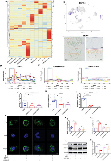
TRPV4 mediated Ca2+ signaling participates the regulation of LIPUS to inflammatory chondrocyte. A) Heatmap showing ion channel gene expression in all cells from the mouse ACLR model of PTOA. B) The express distribution of TRPV4 in UMAP plots. C) Immunohistochemical staining of TRPV4 in the mouse knee joint. Calcium transient relative fluorescence intensity of ROIs with LIPUS stimuli in control group D), in TRPV4‐knockdown group E), and in the TRPV4‐blocked group F). Quantification of the number G) (n = 8), the magnitude H) (n = 8) of calcium peaks, and the responsive rate of cells I) (n = 4). J) Immunofluorescence was used to detect the LC3‐puncta after the chondrocytes was co‐incubated with GSK205. Scale bar, 5 µm. K) Quantification of LC3‐puncta in chondrocytes (n = 4). WB L) and densitometry analysis of Collagen II M) and MMP‐13 N) protein expression of chondrocyte co‐incubated with GSK205 following LIPUS treatment (n = 3). Data are presented as means ± SD. Statistical analysis was performed using Student's t test. ****(P < 0.0001), ***(P < 0.001), **(P < 0.01), *(P < 0.05), ns (0.05 < P).

Expression Data

Expression Detail
Antibody Labeling
Phenotype Data

Phenotype Detail
Acknowledgments
This image is the copyrighted work of the attributed author or publisher, and ZFIN has permission only to display this image to its users. Additional permissions should be obtained from the applicable author or publisher of the image. Full text @ Adv Sci (Weinh)