
Fig. 3 - Supplemental 3
- ID
- ZDB-FIG-250424-44
- Publication
- Campbell et al., 2025 - Opposing roles for Bmp signalling during the development of electrosensory lateral line organs
- Other Figures
- All Figure Page
- Back to All Figure Page
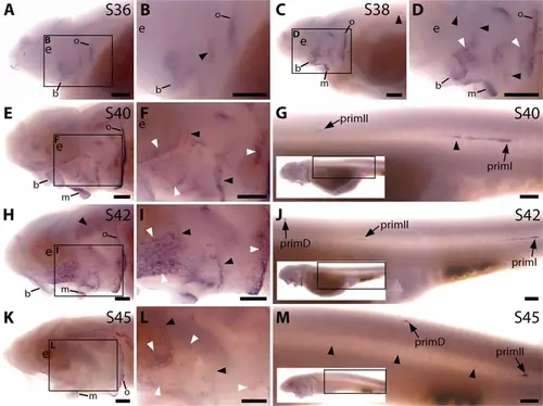
Sterlet Apcdd1 is expressed in ampullary organs and neuromasts during development. In situ hybridisation in sterlet for Apcdd1, encoding a secreted dual Wnt/Bmp antagonist. Black arrowheads indicate examples of developing neuromasts; white arrowheads indicate examples of developing ampullary organs. For trunk images, boxes on low-power insets delineate the regions shown. (A,B) At stage 36, Apcdd1 expression is seen in the region of the preopercular neuromast line, as well as in developing barbel regions, the opercular edge and around the mouth. (C,D) By stage 38, expression in all these locations is more distinct. (E–J) At stage 40 (E–G) and stage 42 (H–J), Apcdd1 is expressed around ampullary organ primordia and neuromasts on the head (E,F,H,I) and is also seen on the trunk in primI and primII and a fairly short line of trailing cells behind primI (G,J). (K–M) At stage 45, this expression pattern largely persists on the head, although appears to be fading in the ventral infraorbital ampullary organ field (K,L). On the trunk, faint expression is seen along the main body line, with strong expression in primD and primII (M). Abbreviations: b, barbels; e, eye; m, mouth; o, operculum edge; prim, migrating lateral line primordium (primI, primary; primII, secondary; primD, dorsal); S, stage. Scale bars: 250 μm. |