
FIGURE
Fig. 1
- ID
- ZDB-FIG-250422-111
- Publication
- Parobková et al., 2025 - Advancing microplastic detection in zebrafish with micro computed tomography: A novel approach to revealing microplastic distribution in organisms
- Other Figures
- All Figure Page
- Back to All Figure Page
Fig. 1
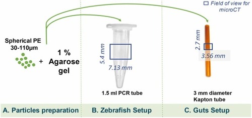
Preparation of reference samples: (A) PE MPs were immersed in agarose gel, (B) transferred into a 1.5 ml PCR tube, chosen to fit within the field of view for the Zebrafish Setup, enabling a voxel size of 4.1 µm. (C) Additionally, the mixture was placed into a 3 mm diameter Kapton tube, fitting the field of view within the Guts Setup. The highlighted fields of view were then scanned using microCT. |
Expression Data
Expression Detail
Antibody Labeling
Phenotype Data
Phenotype Detail
Acknowledgments
This image is the copyrighted work of the attributed author or publisher, and
ZFIN has permission only to display this image to its users.
Additional permissions should be obtained from the applicable author or publisher of the image.
Reprinted from Journal of hazardous materials, 488, Parobková, V., Maleček, L., Zemek, M., Kalčíková, G., Vykypělová, M., Buchtová, M., Adamovský, O., Zikmund, T., Kaiser, J., Advancing microplastic detection in zebrafish with micro computed tomography: A novel approach to revealing microplastic distribution in organisms, 137442137442, Copyright (2025) with permission from Elsevier. Full text @ J. Hazard. Mater.