
Fig. 6
- ID
- ZDB-FIG-250416-37
- Publication
- Le et al., 2025 - Midkine-a interacts with Ptprz1b to regulate neural plate convergence and midline formation in the developing zebrafish hindbrain
- Other Figures
- All Figure Page
- Back to All Figure Page
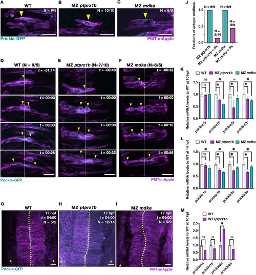
Drosophila Prickle-EGFP rescues midline formation in MZ ptprz1b and MZ mdka mutants that exhibit downregulated prickle expression. (A-C) Representative single-plane images (dorsal views) of neural progenitors in WT (A), MZ ptprz1b (B) and MZ mdka mutant rhombomeres (C) showing localization of injected Drosophila Prickle-EGFP (cyan) at 14 hpf. Cell clones were labelled by co-injection of prickle-EGFP and PMT-mApple mRNA into single cells at the 32-cell stage. Arrowheads indicate anteriorly localized Prickle puncta. Scale bar = 20 μm. (D-F) Representative MIP images of Prickle-EGFP expressing neural progenitors undergoing C-division in WT (D), MZ ptprz1b (E) and MZ mdka mutant rhombomeres (F) between 14 and 17 hpf. Scatter cell labelling was achieved through co-injection of prickle-EGFP and PMT-mApple mRNA into single cells at the 32-cell stage. Time (t) is indicated as hours:minutes (hh:mm). Yellow arrowheads indicate cell postioning during C-division localized. White dashed lines label presumptive midline of neural keel. Scale bar = 20 μm. (G-I) MIP images showing normal midline morphology in rhombomeres at 17 hpf in WT (G), MZ ptprz1b (H) and MZ mdka single mutants (I) injected with prickle-EGFP mRNA. Scale bar = 20 μm. (J) Quantification of ectopic midline penetrance in MZ ptprz1b and MZ mdka mutants injected with PMT-mEGFP/PMT-mApple mRNA with and without prickle-EGFP mRNA. Data for PMT-mEGFP/PMT-mApple mRNA injected mutants were taken from (Fig. 2). Sample numbers are indicated on top of each bar. (K-L) Relative qPCR quantification of prickle1a, prickle1b, prickle2a and prickle2b expression at 10 hpf (K) and 14 hpf (L). Data are shown in the format of mean ± SEM. Asterisks label statistical significance between WT and mutants after statistical analyses (CI = 95%, P < 0.05, see Material and Methods). (M) Relative qPCR quantification of prickle1a, prickle1b, prickle2a and prickle2b expression in WT overexpressing ptprz1b at 10 hpf after injection of ptprz1b-mApple mRNA. Data are shown as mean ± SEM, and statistical significance between WT and ptprz1b-overexpressing WT is labelled by asterisks after statistical analyses (CI = 95%, P < 0.05, see Material and Methods). (For interpretation of the references to colour in this figure legend, the reader is referred to the Web version of this article.) |
Reprinted from Developmental Biology, 521, Le, Y., Rajasekhar, K., Loo, T.Y.J., Saunders, T.E., Wohland, T., Winkler, C., Midkine-a interacts with Ptprz1b to regulate neural plate convergence and midline formation in the developing zebrafish hindbrain, 52-74, Copyright (2025) with permission from Elsevier. Full text @ Dev. Biol.